Prostate cancer is the 2nd most prevalent cancer in men, requiring sophisticated tools for tumor detection and grading, as well as for treatment execution. Recent developments in the field of deep learning and artificial intelligence (AI) are moving the needle in prostate cancer healthcare. More specifically, it is now possible to use state-of-the-art AI and Deep Learning for prostate cancer detection and treatment.
MRI segmentation
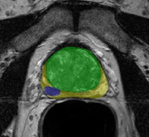
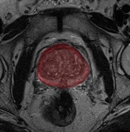
Initial diagnosis of prostate cancer is done using an MRI scan of the gland, and visualization of the suspected areas. This process is rather challenging as it is highly dependent on the radiologist’s image interpretation and quality of scan. Deep learning technology can be used to perform prostate gland segmentation on MRI scans, assess tumor risk and develop automated measurements for Gleason Score. These can be done accurately and precisely, therefore reducing the need for invasive procedures (biopsy) and providing a baseline segmentation. This segmentation can be used by clinicians to locate the areas of risk within the gland and suggest a roadmap for biopsies.
Advanced deep learning algorithms are proven to work well for various medical tasks, and prostate gland segmentation is no exception. Deep neural network architectures that are built for the segmentation task, and trained on prostate gland data, produce very accurate results for further analysis. Classical algorithms are struggling with the complex textures of the prostate gland, and often challenging cases do not work well. Deep learning uses data to build internal features of the gland. This increases success rate on a variety of images, including very challenging ones.
Ultrasound segmentation, 2D to 3D, and registration
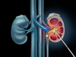
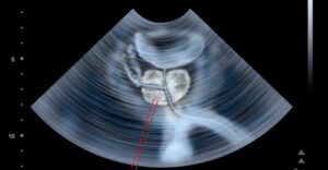
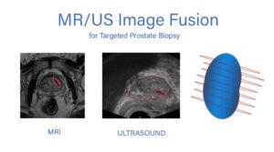
During a biopsy, a needle is guided by ultrasound imaging, sampling various areas of the gland (specifically those previously defined as suspicious) based on the MRI scan. However, ultrasound guidance of the needle is intricate, as ultrasound images are limited in quality. Characteristic images have significant noise level, depending on the position of the ultrasound probe, and the tissue properties between the probe and the target organ. In addition, interpreting the image is highly dependent on the user, suggesting that an objective source for biopsy guidance will increase the success of the procedure by verifying adequate scatter of the samples from the prostate gland, and higher accuracy for sampling of the pathologies within the gland. Transforming the ultrasound 2D images into a 3D model of the prostate gland is feasible using advanced AI algorithms. This allows spatial view of the gland to improve navigation by adding a depth dimension to the procedure, and assisting with precise needle positioning. Furthermore, registration (or fusion) of the ultrasound image with the MRI scan can provide the clinician with a “map” of the gland, while the real-time ultrasound images are accurately fused with it. This utilizes the superior resolution and quality of the MRI images, in contrast to the ultrasound images, thus allowing for easier navigation. This significantly shortens procedure length and reduces complications.
Automated microscopy analysis
Personalized treatment requires microscopic analysis of biomarkers within the patient’s tissue sample. Current protocol requires manual assessment of image fluorescence (IF), a time-consuming task. Using an automated system for image fluorescence quantification is comparable to human analysis and reduces manual labor.
Robotic guidance in procedures
Artificial Intelligence and deep learning systems have been proven to assist in minimal-invasive robotic surgeries, specifically in robotic guidance and feedback, by powering multiple tools such as registration, segmentation and tracking. Prostatectomy, a common treatment of prostate cancer, can benefit from the use of these advanced algorithms to increase procedural success. Tool tracking and guidance will lower complications rate, since tracked tools can be activated only if they are properly visible and are in the correct place. These can be achieved using real-time imaging analysis.
RSIP Vision’s work in Deep Learning for Prostate Cancer
As the use of imaging modalities for prostate cancer treatment increases, so does the need for advanced image analysis techniques. AI and deep learning custom algorithms can be integrated into all steps of prostate cancer care, from detection to treatment, thus improving patient outcome. RSIP Vision’s experience and expertise, developing such capabilities in medical devices and applications, can be used to implement these algorithms into a variety of devices and solutions, enhancing clinical care and shortening time to market.
 Urology