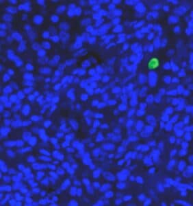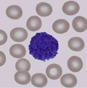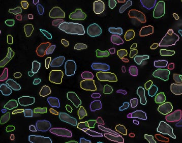
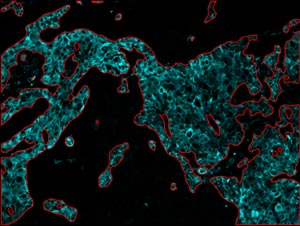

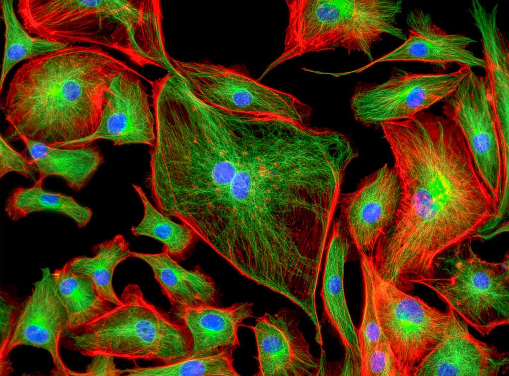
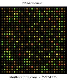

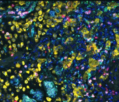


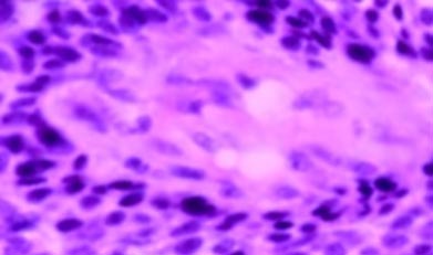

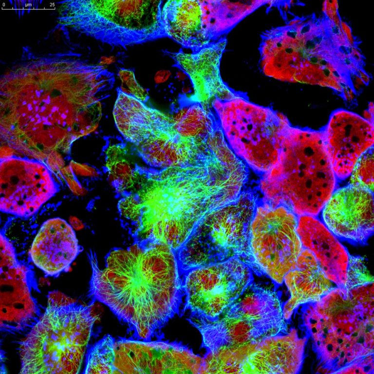
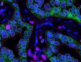
Multiplex IF Analysis
The use of deep learning for analysis of multiplex IF has allowed for a much greater accuracy level for the correct phenotypic classification of cells. When combined with RSIP Vision‘s advanced nuclear detection capability, it allows for the simultaneous analysis of multiple florescent markers on a cell by cell basis. This tool is well suited for multiple applications, especially when using multiple markers to characterize distinct cell populations such as in immune-oncology and IBD.