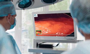


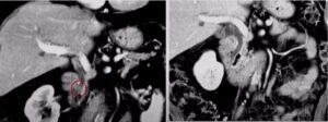
Image Analysis and AI in Diagnostic ERCP

Enhanced ERCP tumor assessment using AI
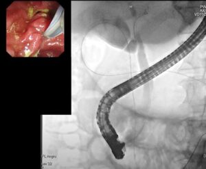
AI in ERCP gallstones and strictures treatment
AI for Gastric Cancer Detection
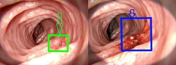
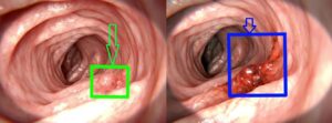
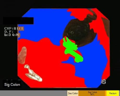
Upper gastrointestinal cancers, including esophageal cancer and gastric cancer, are among the most common cancers worldwide. However, a lack of endoscopists with colonoscopy skills has been identified and solutions are critically needed. The development of a real-time robust detection system for colorectal neoplasms is needed to significantly reduce the risk of missed lesions during colonoscopy.
Gastrointestinal lesion detection with machine learning
Image Analysis and AI in Diagnostic ERCP
Enhanced ERCP tumor assessment using AI
AI in ERCP gallstones and strictures treatment
AI for Gastric Cancer Detection
Upper gastrointestinal cancers, including esophageal cancer and gastric cancer, are among the most common cancers worldwide. However, a lack of endoscopists with colonoscopy skills has been identified and solutions are critically needed. The development of a real-time robust detection system for colorectal neoplasms is needed to significantly reduce the risk of missed lesions during colonoscopy.