
 Fully automatic
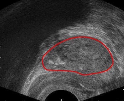
PRESS RELEASE – RSIP Vision introduces an innovative set of AI modules for enhanced medical ultrasound applications
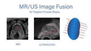
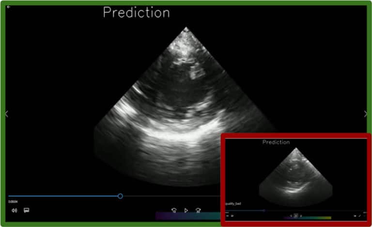
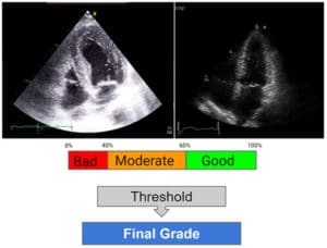
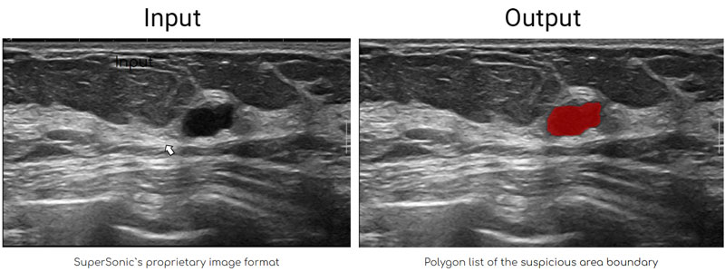
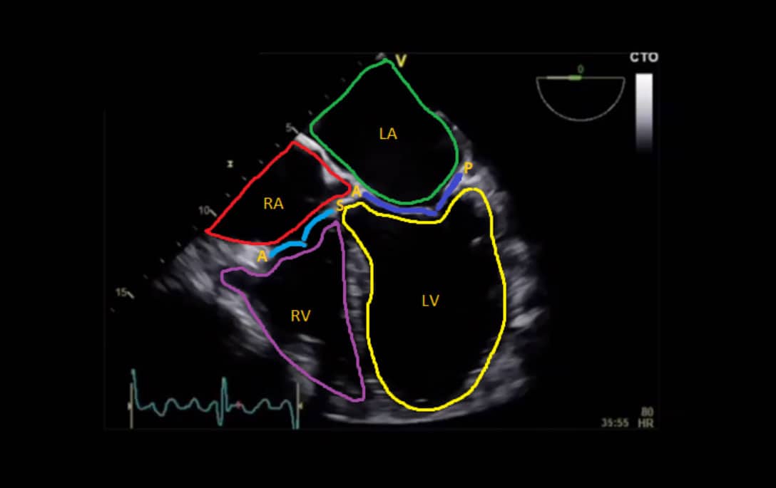
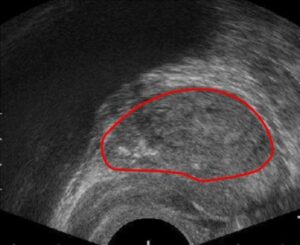
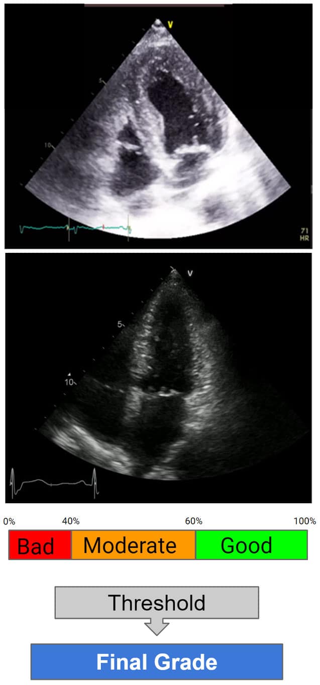
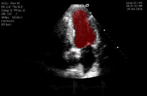
RSIP Vision introduces an innovative set of AI modules for enhanced medical ultrasound applications. These innovative modules empower a wide range of medical applications by overcoming the main ultrasound challenges – user-dependent acquisition and noisy, clinically challenging images. This improves the workflow and diagnostic accuracy while reducing the overall procedure time. SILICON VALLEY, Calif., June 22, 2020 — RSIP Vision, a global leader in artificial intelligence (AI) and computer vision technology, announced today a new set of AI-based medical ultrasound modules. These advanced modules will serve as AI-based building blocks