


 Deep-learning based analysis
 Deep-learning based analysis

PR – Non-Invasive Planning of Coronary Intervention
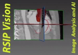
RSIP Vision Presents New Technology for Non-Invasive Planning of Coronary Intervention Innovative technology provides accurate coronary artery 3D reconstruction from 2D angiography to be used for diagnosis, measurements, stent modelling, and characterization for procedural planning. TEL AVIV, Israel & SAN JOSE, Calif., December 8, 2021 – RSIP Vision, an experienced leader in driving innovation for medical imaging through advanced AI and computer vision solutions, today announces a new coronary artery modelling technology. This technology enables quick and accurate reconstruction of the coronary vasculature during angiography into a 3D model. This