More than 20,000 adults in the United States (with a higher incidence on men vs. women) are diagnosed every year with primary cancerous tumors of the brain and the spinal cord. It is estimated that more than 15,000 die from this disease yearly. An additional 4,000 children and teens are diagnosed with a brain or central nervous system tumor. In addition to primary tumors, the brain can also suffer from secondary tumors or brain metastases. The most common cancers that spread from remote areas to the brain are lung, breast, melanoma, kidney, nasal cavity and colon cancers.
The task of detecting the localization and extension of the tumor falls on brain tumor image processing, by the way of segmenting the tumor in the image. Segmentation of anatomical structure is a challenging assignment. The variability in target structures limits the use of a-prior knowledge which would restrict the expected geometry of the object. In particular, anatomical structure with unexpected shape, like soft tissue tumors, challenges automatic segmentation and in most parts requires human supervision to complete segmentation.
The task of detecting the localization and extension of the tumor falls on brain tumor image processing, by the way of segmenting the tumor in the image. Segmentation of anatomical structure is a challenging assignment. The variability in target structures limits the use of a-prior knowledge which would restrict the expected geometry of the object. In particular, anatomical structure with unexpected shape, like soft tissue tumors, challenges automatic segmentation and in most parts requires human supervision to complete segmentation.
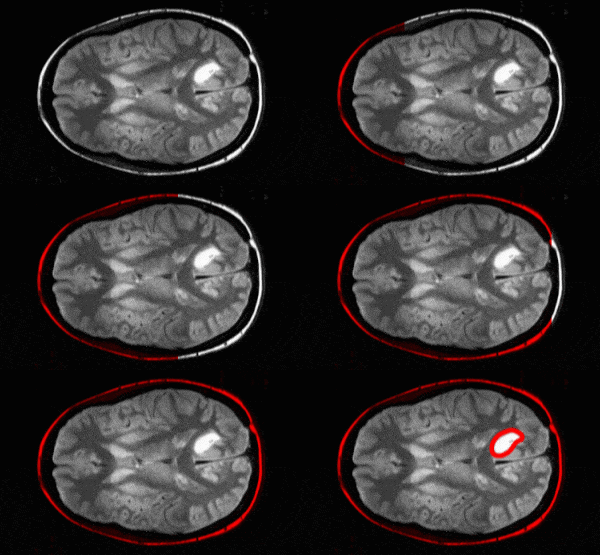
Three-dimensional segmentation of brain tumor has an important clinical relevance for the estimation of the volume and spread of the tumor. The first step in segmentation is to construct a probability map localizing the tumor. A binary fuzzy classification of voxels based on intensity histogram into tumor and background classes is employed for the task.
Using the probabilistic maps, we can employ deformable models – like active contours (snakes) – to delineate the tumor. Active contours are parametric description of a closed curve, made to converge to regions of the image corresponding to edges. Thus, the probability maps are used to initialize and guide the convergence of the active contour to the expected tumor boundaries.
Convergence of level set active contours is the core of successful brain tumor segmentation. A minimization of an energy functional defining the active contour is made in high dimensional domain, regarding which the object boundaries correspond to the zero level energy. A correct definition of the active contour is an art by itself. This is specifically true about the energy functional and the image values it relies upon.
At RSIP Vision we have already employed active contour segmentation in medical applications for a full decade. The expertise we have accumulated allows us to develop cutting edge solutions for segmentation in low signal to noise ratio and in complex environments as well.

