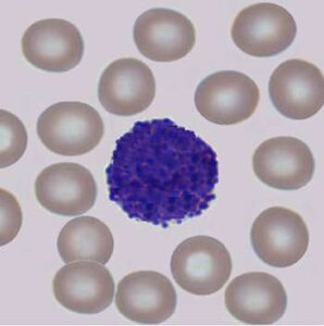
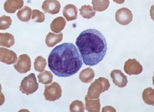
Circulating tumor cells (CTCs) are rare cancer cells that originate from a tumor and then travel through the patient’s blood or lymphatic system. CTCs have a high risk of metastasizing elsewhere in the body, which can be very dangerous. It is well-known that finding and analyzing these rare cells can give a very good basis for the right prognosis and the most suitable treatment for the patient.
The problem with CTCs is that it is very difficult to find them among thousands of regular cells. This makes testing for them quite expensive. Developments in AI enable us to do this tedious manual work much more efficiently by detecting the CTCs on slides by screening out the regular cells.
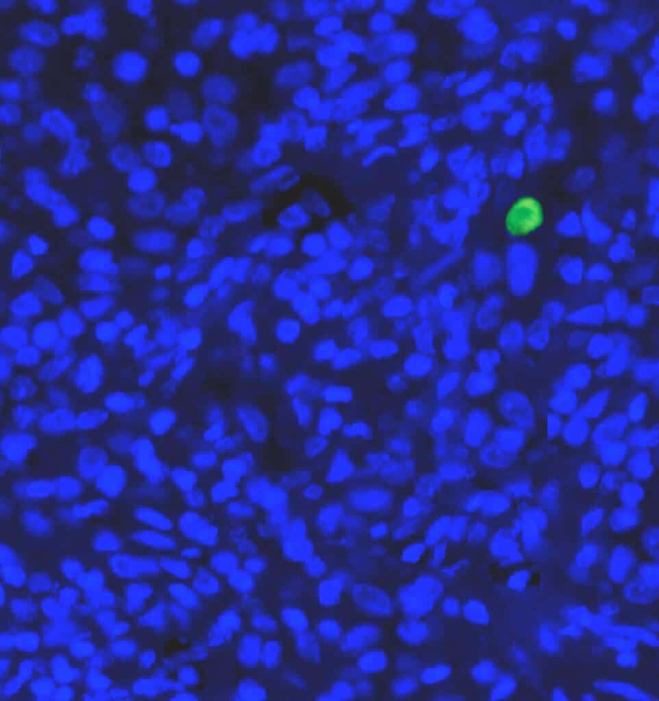
Automated CTCs detection
From RSIP Vision’s experience, we would like to share some insights on how to solve this problem. First and most importantly is to train the AI algorithm, which is based on a convolutional neural network, with the rare cells. Of course, the training is inherently imbalanced because there are many more healthy cells than cancerous ones, making the task very difficult. To compensate for the discrepancy in numbers, data augmentation must be used to produce more possible appearances of CTCs that will enable the system to learn to do this distinction.
The second insight is to be prepared for several changes during the development cycle of the algorithm, since the data poverty of positive cells is likely to turn into a multi-phase-long period of data collection. In real life this translates to version one, then version two, and so forth. Between each version the data can change the algorithm considerably, so make sure to budget sufficient resources for the entire run of iterations.
The last tip concerns data annotation. You can show a false negative and a false positive to a cytologist, and in many cases they can explain and guide your next level of development. In some cases they will change the annotations and in others they will point out a third category of cells, which is important to use when training the algorithm. As usual, it is essential to be fully aligned with the clinician or the pathologist.
Nowadays there is an industry-wide effort to enable detection of CTCs because it is a common belief that such tests can help gain insight into different processes in the development of cancer. RSIP Vision is using its algorithm and image analysis expertise to pioneer the way to automated CTC detection.
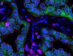
 Microscopy
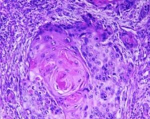