Urolithiasis Healthcare Using AI
Urolithiasis, the formation of “stones” in the urinary pathway, is a prevalent pathology, found in 4-20% of the population. Recent developments in the field of image analysis, specifically deep learning (DL) and artificial intelligence (AI) are improving urolithiasis detection and treatment and enhancing the clinical outcome.
Deep Learning in stone detection and classification
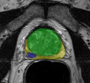
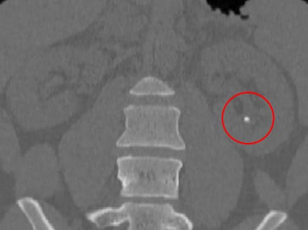
Initial detection of urolithiasis requires ultrasound or CT imaging. Identification of all the stones and their position can be challenging. Numerous stones can exist simultaneously and can be easily overlooked, even by experienced physicians. Utilizing convolutional neural networks (CNN), trained and adapted to suit this ultrasound and CT data, results in accurate stone recognition and segmentation, thus reducing manual labor, and speeding the image analysis process. Since these CNN allow a pixel-level analysis of the images and image enhancement techniques, they enable the use of lower-quality CT images, therefore reducing radiation-dosage during the scan.
Segmentation of stones can be performed using neural networks. This task is not straightforward as stones take a very small portion of the image. To this end, a common U-Net architecture, that was shown to work well on various medical segmentation tasks, was adapted to segment the small stones. This was done using a special loss function, which focuses mostly on stones’ neighborhood.
Understanding the stone-type is essential for determining the optimal treatment. Up-to-date methods for stone classification are based on urine tests which are time-consuming or a painful wait for spontaneous stone passing. The suggested segmentation network can be enhanced to jointly produce both segmentation and stone type. By solving both tasks simultaneously, stone type classification can benefit from the network’s knowledge on the location of the stone. Kidney stone CT data can classify the stones into their subtypes with notable accuracy, assisting and speeding treatment selection.
Deep Learning in urolithiasis treatment
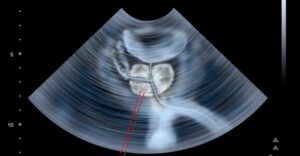
Shockwave lithotripsy
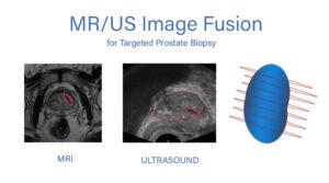
Shockwave lithotripsy (SWL) requires precise aim of the wave’s focus and is performed under x-ray guidance. Misdirection of the focus could result in kidney damage and procedural failure. Implementing Deep Learning algorithms to fuse the x-ray images with the preprocedural CT scan allows a 3D view of the anatomy, improving SWL accuracy, and increasing procedural success. For this task, incorporating deep learning segmentation with classical optimization techniques that find the optimal transform between the two, yields a very accurate result. A “simulated X-ray image” can be produced out the CT and an estimated X-ray emitter location. This location can be optimized to produce an image that is as close as possible to the actual X-ray image, a task which can be performed using the cycle GAN framework. At this stage, both modalities can be merged. In addition, the x-ray images can be rendered to a 3D model, another procedural aid to ease shockwave focus aim.
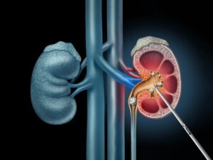
Percutaneous Nephrolithotomy or Nephrolithotripsy (laser treatment)
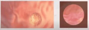
Percutaneous solutions are guided by real-time cameras. Additional guidance for laser aim or stone breaking could reduce complications. Tracking of the equipment using both hardware upgrades and state-of-the-art CNN algorithms, as well as 3D reconstruction of the images, allows easier and more accurate performance during the procedure, improving stone breaking results, as well as better placement of stents if needed.
Stone passing
Often the stones will pass naturally. Determining the potential of a stone to pass naturally could prevent unnecessary interventions. Deep Learning algorithms can segment the various organs and extract properties of each. Post processing that extracts the optimal path and distance from boundaries in the ureter segmentation can be used to calculate the route a stone needs to travel for spontaneous passage and determine if a stone can pass freely through it. Classification of these stones can separate them from those who need intervention. This will shorten procedure time and notify the physician regarding areas of concern.
Post-op care with image analysis
Defining procedural success is challenging. The physician must determine if the removed stones are sufficient for symptom relief, or if further treatment is required. Comparing the images post-op, or even during the procedure, to the baseline scan can alert of upcoming pathologies. Stone segmentation and precise image registration compare the post-op state to the pre-op pathology and detect stones that still require immediate removal, debris that may obstruct urological canals, and misplaced stents. Rather than waiting for the next complication, using these AI techniques, may prevent it.
RSIP Vision improves urolithiasis healthcare using AI
Throughout the full cycle of detection and treatment of urolithiasis, multimodal imaging techniques are involved. Utilizing RSIP Vision’s vast experience in developing custom Deep Learning and AI image analysis algorithms in the medical field can significantly improve urolithiasis outcome. We understand that each device has its unique challenges, and as we develop customer-specific solutions, we can help in building the future solutions in the field of urolithiasis healthcare with AI.
 Urology