Skip to content
  • Our Work
    • Fields
      • Cardiology
      • ENT
      • Gastro
      • Orthopedics
      • Ophthalmology
      • Pulmonology
      • Surgical Intelligence
      • Surgical Robotics
      • Urology
      • Other
    • Modalities
      • Endoscopy
      • Medical Segmentation
      • Microscopy
      • Ultrasound
  • Success Stories
  • Insights
    • Magazine
    • Upcoming Events
    • Webinars
    • Meetups
    • News
    • Blog
  • The company
    • About us
    • Careers
Menu
  • Our Work
    • Fields
      • Cardiology
      • ENT
      • Gastro
      • Orthopedics
      • Ophthalmology
      • Pulmonology
      • Surgical Intelligence
      • Surgical Robotics
      • Urology
      • Other
    • Modalities
      • Endoscopy
      • Medical Segmentation
      • Microscopy
      • Ultrasound
  • Success Stories
  • Insights
    • Magazine
    • Upcoming Events
    • Webinars
    • Meetups
    • News
    • Blog
  • The company
    • About us
    • Careers
Contact

Lymph Node Segmentation Module

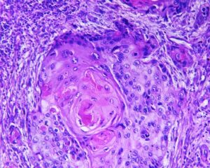
Lymph nodes are routinely examined and assessed during physical examination of patients in a clinic or hospital setting. Enlarged lymph nodes can be indicators of infection, cancer and other pathologies. Therefore, a biopsy of a suspected lymph node, which provides tissue histology or cytology is a vital step towards diagnosis.

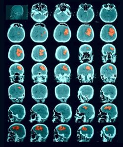
In relation to malignancy, initial staging before treatment is done using the Ann Arbor staging system or Lugano classification, by analyzing patient’s computed tomography (CT) and positron emission tomography (PET) scans. In addition, tracking the changes in lymph node’s size, is indicated during and after treatment e.g, radiation or chemotherapy. This monitoring may reflect treatment effectiveness. Again, the assessment of response can be done using the Lugano classification, and more recently by the lymphoma response to immunomodulatory therapy criteria (LYRIC).

Automated lymph node segmentation

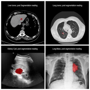
Analysis of lymph nodes can be difficult and time-consuming for the radiologist. Hence, automatic detection and segmentation of lymph nodes from CT scans is important and valuable in order to eliminate errors. Automated lymph nodes segmentation requires expertise in differentiating lymph node boundaries from adjacent structures. An automatic algorithm would provide the physician with relevant information for and during the lymph node biopsy and also supply accurate information for diagnosis and management.

RSIP Vision built an advanced AI module, based on the cutting edge convolutional neural network architecture, for segmentation of lymph nodes in CT scans using deep learning algorithms. This module was fine tuned to withstand the various challenges encountered while trying to determine whether each voxel belongs to a lymph node or not and resulted in one of the quickest and most accurate ways to perform this task. If you are still performing this task manually, we have the ideal software solution for you. Talk to an RSIP Vision engineer now.

Share

Share on linkedin
Share on twitter
Share on facebook

Main Field

Medical segmentation

RSIP Vision is very active in all fields of medical image processing and computer vision applications. Besides all our work in the domain of Artificial Intelligence for cardiology, ophthalmology, pulmonology and orthopedics, our engineers have contributed to many other medical segmentation projects helping our clients to improve public health and save thousands of lives. These medical applications in computer vision help physicians perform early identification of major diseases in brain, kidney, prostate and many other organs. Contact us and tell us about your medical computer vision project: we will help you complete with success all medical segmentation tasks.

View Medical segmentation

Categories

  • Medical segmentation, RSIP Vision Learns

Related Content

one-click segmentation

One-click segmentation of medical images

Liver and tumors

Liver Tumor Segmentation with Deep Learning

Lesion Detection in CT scan (Hemorrhagic stroke)

Lesion segmentation by random-forest classifiers

Squamous cell carcinoma

Automatic segmentation of tumor cells

one-click segmentation

One-click segmentation of medical images

Liver and tumors

Liver Tumor Segmentation with Deep Learning

Lesion Detection in CT scan (Hemorrhagic stroke)

Lesion segmentation by random-forest classifiers

Squamous cell carcinoma

Automatic segmentation of tumor cells

Show all

RSIP Vision

Field-tested software solutions and custom R&D, to power your next medical products with innovative AI and image analysis capabilities.

Read more about us

Get in touch

Please fill the following form and our experts will be happy to reply to you soon

Recent News

PR – Intra-op Virtual Measurements in Laparoscopic and Robotic-Assisted Surgeries

PR – Non-Invasive Planning of Coronary Intervention

PR – Bladder Panorama Generator and Sparse Reconstruction Tool

PR – Registration Module for Orthopedic Surgery

All news
Upcoming Events
Stay informed for our next events
Subscribe to Our Magazines

Subscribe now and receive the Computer Vision News Magazine every month to your mailbox

 
Subscribe for free
Follow us
Linkedin Twitter Facebook Youtube

contact@rsipvision.com

Terms of Use

Privacy Policy

© All rights reserved to RSIP Vision 2021

Created by Shmulik

  • Our Work
    • title-1
      • Ophthalmology
      • Uncategorized
      • Ophthalmology
      • Pulmonology
      • Cardiology
      • Orthopedics
    • Title-2
      • Orthopedics
  • Success Stories
  • Insights
  • The company