RSIP Vision Announces Sophisticated AI-Based Tool for Coronary Artery Analysis and Intervention Planning
New module utilizes state-of-the-art deep learning algorithms combined with classic computer vision methods to create a 3D model of the coronary arteries to better visualize artery structure and reduce procedural risks to patients.
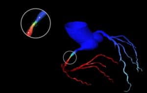
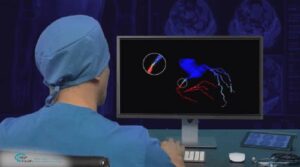
TEL AVIV, Israel & SAN JOSE, Calif., February 16, 2021 – RSIP Vision, an experienced leader in driving innovation for medical imaging through advanced AI and computer vision solutions, today announces a new coronary artery segmentation tool. The fully automated, deep learning-based technology provides a quick, robust, and accurate 3D model of coronary artery anatomy, enabling precise measurements of the artery length and diameter at any point. This tool enhances image-based assessments of coronary artery function and diagnosis of conditions, such as narrowing (stenosis). It also provides the needed understanding to physicians for advanced procedure planning, including navigation planning, stent selection, and stent positioning. Other benefits include:
.
- Reduced procedural risks to the patient
- Comprehensive 3D modeling of all vessels
- Shorter procedure time
- Increase ability to make quick and accurate clinical decisions
“The cardiac CT-scan contains vast amounts of data surrounding the coronary arteries, and our mission is to extract it,” said Ron Soferman, founder & CEO at RSIP Vision. “Its wide availability and excellent resolution make it a preferred imaging modality for cardiology purposes. We are pleased to introduce our new module that will aid in the detection of coronary artery stenosis and be useful in percutaneous coronary intervention (PCI) planning. We believe strongly this will reduce the need for additional interventions, while decreasing the chance of adverse events due to incorrect stent selection.”
This new module was designed to better visualize artery structure, measure the vessel dimensions in points-of-interest, and enable accurate, image-based FFR measurements. Additional modules can show artery modification due to stent placement or place a virtual stent in the desired position within the coronary artery. These uses replace invasive cardiac measurements and give the physician a better pre-procedure planning tool for stent selection.
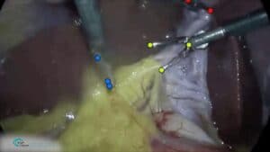
“Proper pre-procedural planning is paramount for any invasive cardiac intervention, especially in PCI,” said Dr. David Yakobi, Board Certified Cardiac Surgeon. “Any automated technology that eliminates human error and bias contributors has the potential to augment procedural success, increase stent patency rate in the long-run and avoid unnecessary consecutive interventions. This module from RSIP Vision will also allow us to integrate its analytical findings into the SYNTAX Score and classify the severity of patients’ combined coronary stenoses so the interventional cardiologist can predict the outcome of the procedure according to anatomical and pathological variables to guide the choice between PCI and CABG (Coronary Artery Bypass Surgery) and predict long-term survival.”
Consistent with RSIP Vision’s preceding, reliable solutions, this new tool is vendor-neutral and available to third-party medical device and application vendors, as well as CT manufacturers. The initial version will provide coronary arteries segmentation in contrast CT scans. Going forward, RSIP Vision plans to extend additional functionalities including calcification detection, support for pre-existing stents, and 3D reconstruction from 2-dimensional angiography.

