Skip to content
  • Our Work
    • Fields
      • Cardiology
      • ENT
      • Gastro
      • Orthopedics
      • Ophthalmology
      • Pulmonology
      • Surgical Intelligence
      • Surgical Robotics
      • Urology
      • Other
    • Modalities
      • Endoscopy
      • Medical Segmentation
      • Microscopy
      • Ultrasound
  • Success Stories
  • Insights
    • Magazine
    • Upcoming Events
    • Webinars
    • Meetups
    • News
    • Blog
  • The company
    • About us
    • Careers
Menu
  • Our Work
    • Fields
      • Cardiology
      • ENT
      • Gastro
      • Orthopedics
      • Ophthalmology
      • Pulmonology
      • Surgical Intelligence
      • Surgical Robotics
      • Urology
      • Other
    • Modalities
      • Endoscopy
      • Medical Segmentation
      • Microscopy
      • Ultrasound
  • Success Stories
  • Insights
    • Magazine
    • Upcoming Events
    • Webinars
    • Meetups
    • News
    • Blog
  • The company
    • About us
    • Careers
Contact

Three-dimensional reconstruction of a deformable object

Reconstruction of the three-dimensional surface of an object based on single view 2-D sequence of images is a highly challenging task. Challenges stem in part from the construction of a template representing the object, or more formally, incorporating knowledge to restrict the shape space. The possible deformation of the object needs to be known in advance in order to be able to match it to the temporal information coming from a sequence of images or a dynamic point cloud. The end result of deformable object reconstruction is a surface and a temporal deformation model.
CT scan of healthy lungs

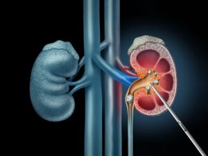
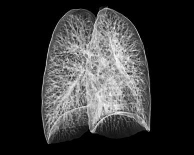
Applications of deformable object reconstruction can be found in several fields: one of those is clinics, where monitoring the state of an organ by CT, angiography or x-ray can provide important prognostic insight before invasive procedure are engaged. Furthermore, once a candidate surface has been reconstructed to represent the target organ, a virtual model can be used for real-time navigation, where catheters or other equipment is tracked and its 3D position is overlaid on the reconstructed model. Physicians can then navigate in an otherwise occluded environment inside the body, increasing operation accuracy and possibly preventing injuries caused by equipment manipulation.

Three-dimensional reconstruction of an object

The three-dimensional reconstruction of a deformable object includes two main parts. The first involves tracking, feature matching or the establishment of temporal link between successive points on the surface. The second part includes surface representation of the target object. The trajectories tracked can be used for learning the temporal relationship between the set of points, which produces a mathematical representation of their motion. The importance of learning these trajectories lies in the ability to put them into use for motion predictor-correctors systems such as the Kalman filters. Doing so, we can pose restriction on the global motion (envelope) deformation of the object. This dynamic point cloud is then used for surface fitting. The surface representing the object should be flexible to take many forms, but not violate too strongly the deformation restrictions. Surface matching smooths out irregularities in the point cloud and enables a clearer view of the object.

The model to be fitted to the object is oftentimes learned from a template database, which is constructed offline. Using an object database, multiple templates can be created, enabling us to learn the shape space. Furthermore, the template database allows us to extract features related to object’s appearance and its deformation constraints. This knowledge can be put to use when reconstructing an object in an occluded environment, where, as is many times the case, we have to isolate the target object from its surrounding.

The technology leading to surface reconstruction of rigid objects with predictable shape from a sequence of images is quite advanced. However, this is not the same for deformable objects. The three dimensional reconstruction of an object demands a careful construction of the deformation shape space and an accurate mathematical model to represent its motion. Additional steps involving feature extraction and image pre-processing are essential to assure correct surface representation. These challenges are yet to be solved with an off-the-shelf technique and therefore require custom development.

RSIP Vision specializes in constructing tailor-made algorithmic solutions for image processing, computer vision and machine learning tasks. We have developed the expertise to handle reconstruction challenges from data acquisition to 3D representation. You will find in RSIP Vision’s project page much more information about our customized solutions, including our work and research in 3D reconstruction.

Share

Share on linkedin
Share on twitter
Share on facebook

Related Content

Percutaneous Nephrolithotomy

PCNL – Planning and real-time navigation

Prostate Tumor Segmentation

Implementing AI to Improve PI-RADS Scoring

RAS Navigation

Tissue Sparing in Robotic Assisted Orthopedic Surgeries

Procedural Planning in urology

Procedural Planning in Urology

C Arm X-Ray Machine Scanner

Radiation Reduction in Robotic Assisted Surgeries (RAS) Using AI

Visible spectrum color

Hyperspectral Imaging for Robotic Assisted Surgery

Show all

RSIP Vision

Field-tested software solutions and custom R&D, to power your next medical products with innovative AI and image analysis capabilities.

Read more about us

Get in touch

Please fill the following form and our experts will be happy to reply to you soon

Recent News

PR – Intra-op Virtual Measurements in Laparoscopic and Robotic-Assisted Surgeries

PR – Non-Invasive Planning of Coronary Intervention

PR – Bladder Panorama Generator and Sparse Reconstruction Tool

PR – Registration Module for Orthopedic Surgery

All news
Upcoming Events
Stay informed for our next events
Subscribe to Our Magazines

Subscribe now and receive the Computer Vision News Magazine every month to your mailbox

 
Subscribe for free
Follow us
Linkedin Twitter Facebook Youtube

contact@rsipvision.com

Terms of Use

Privacy Policy

© All rights reserved to RSIP Vision 2021

Created by Shmulik

  • Our Work
    • title-1
      • Ophthalmology
      • Uncategorized
      • Ophthalmology
      • Pulmonology
      • Cardiology
      • Orthopedics
    • Title-2
      • Orthopedics
  • Success Stories
  • Insights
  • The company