Skip to content
  • Our Work
    • Fields
      • Cardiology
      • ENT
      • Gastro
      • Orthopedics
      • Ophthalmology
      • Pulmonology
      • Surgical Intelligence
      • Surgical Robotics
      • Urology
      • Other
    • Modalities
      • Endoscopy
      • Medical Segmentation
      • Microscopy
      • Ultrasound
  • Success Stories
  • Insights
    • Magazine
    • Upcoming Events
    • Webinars
    • Meetups
    • News
    • Blog
  • The company
    • About us
    • Careers
Menu
  • Our Work
    • Fields
      • Cardiology
      • ENT
      • Gastro
      • Orthopedics
      • Ophthalmology
      • Pulmonology
      • Surgical Intelligence
      • Surgical Robotics
      • Urology
      • Other
    • Modalities
      • Endoscopy
      • Medical Segmentation
      • Microscopy
      • Ultrasound
  • Success Stories
  • Insights
    • Magazine
    • Upcoming Events
    • Webinars
    • Meetups
    • News
    • Blog
  • The company
    • About us
    • Careers
Contact

Endoscopic Procedures using Intravenous OCT

While ultrasound imaging cannot be replaced, adding Intravenous OCT to endoscopy procedures significantly improves image resolution and increases the ability to detect plaque and segment it.

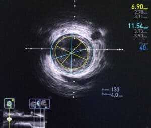
Intravenous Optical Coherence Tomography (IVOCT) is a catheter-based medical procedure which uses a near infra-red light to examine the invitro vascular wall structure. Its ability to reach high-resolution – around 10 microns – means it is able to detect very thin layers of plaque in veins, layers which the 150-200 micron resolution of regular ultrasound usually misses. Images acquired using intravenous OCT, have been analyzed successfully using classical techniques such as support-vector machines. However, deep learning and convolutional neural networks (CNN) are required to get the most out of these images; with them, accuracy of over 90 per cent can be achieved.

Intravenous OCT

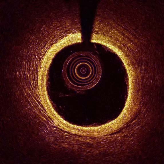
Despite its high resolution, when comparing penetration depth intravenous OCT, which has only a 1-2-millimeter penetration range, falls short of ultrasound. Each imaging modality has different strengths, and best results are achieved when using a combination of both. We maximize our detection capabilities by using image registration to align the two scans so that they represent the same exact area and position. From there a neural network analyzes each image offline and uses the best results from each.

Ilya Kovler CTO at RSIP Vision adds: “From my experience, the high degree of speckle noise in OCT images can hamper the plaque segmentation task. Therefore, adding a pre-processing stage to reduce this noise is critical to the success of the segmentation.”
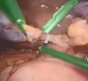
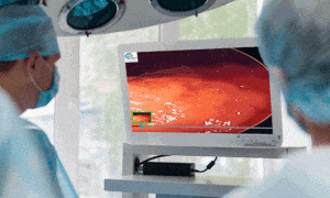
Share

Share on linkedin
Share on twitter
Share on facebook

Main Field

Endoscopy

AI in Endoscopy is a new field, utilizing innovative AI technologies to enhance the accuracy of Endoscopy imaging and provide better detection and clinical outcomes. RSIP Vision is a pioneer in the field of artificial intelligence for endoscopy imaging. Thanks to our deep learning solutions, radiologists and physicians obtain much more information than before from endoscopy images for both detection of diseases and measurement of therapy progress effectiveness. We have successfully completed a large number of important R&D projects for all kind of minimally invasive procedures. Contact us and discuss your project with our experts!

View Endoscopy

Categories

  • Endoscopy, RSIP Vision Learns

Related Content

Healthy gut - 3D Rendering

Real time SLAM in Endoscopy Applications

Intravenous Ultrasound

Using AI to Analyse Intravenous Ultrasound Images

Surgical Instrument Segmentation

Surgical Workflow Analysis

AI-based Surgical Workflow Analysis, another big step towards the future of robotic surgery

Healthy gut - 3D Rendering

Real time SLAM in Endoscopy Applications

Intravenous Ultrasound

Using AI to Analyse Intravenous Ultrasound Images

Surgical Instrument Segmentation

Surgical Workflow Analysis

AI-based Surgical Workflow Analysis, another big step towards the future of robotic surgery

Show all

RSIP Vision

Field-tested software solutions and custom R&D, to power your next medical products with innovative AI and image analysis capabilities.

Read more about us

Get in touch

Please fill the following form and our experts will be happy to reply to you soon

Recent News

PR – Intra-op Virtual Measurements in Laparoscopic and Robotic-Assisted Surgeries

PR – Non-Invasive Planning of Coronary Intervention

PR – Bladder Panorama Generator and Sparse Reconstruction Tool

PR – Registration Module for Orthopedic Surgery

All news
Upcoming Events
Stay informed for our next events
Subscribe to Our Magazines

Subscribe now and receive the Computer Vision News Magazine every month to your mailbox

 
Subscribe for free
Follow us
Linkedin Twitter Facebook Youtube

contact@rsipvision.com

Terms of Use

Privacy Policy

© All rights reserved to RSIP Vision 2021

Created by Shmulik

  • Our Work
    • title-1
      • Ophthalmology
      • Uncategorized
      • Ophthalmology
      • Pulmonology
      • Cardiology
      • Orthopedics
    • Title-2
      • Orthopedics
  • Success Stories
  • Insights
  • The company