Skip to content
  • Our Work
    • Fields
      • Cardiology
      • ENT
      • Gastro
      • Orthopedics
      • Ophthalmology
      • Pulmonology
      • Surgical Intelligence
      • Surgical Robotics
      • Urology
      • Other
    • Modalities
      • Endoscopy
      • Medical Segmentation
      • Microscopy
      • Ultrasound
  • Success Stories
  • Insights
    • Magazine
    • Upcoming Events
    • Webinars
    • Meetups
    • News
    • Blog
  • The company
    • About us
    • Careers
Menu
  • Our Work
    • Fields
      • Cardiology
      • ENT
      • Gastro
      • Orthopedics
      • Ophthalmology
      • Pulmonology
      • Surgical Intelligence
      • Surgical Robotics
      • Urology
      • Other
    • Modalities
      • Endoscopy
      • Medical Segmentation
      • Microscopy
      • Ultrasound
  • Success Stories
  • Insights
    • Magazine
    • Upcoming Events
    • Webinars
    • Meetups
    • News
    • Blog
  • The company
    • About us
    • Careers
Contact

Using AI to Analyse Intravenous Ultrasound Images

While the low-resolution Intravenous Ultrasound (IVUS) present its challenges, by adding AI to the we can easily overcome this issue, and improve the analysis of blood vessels interior parts for a better clinical outcome.

Background

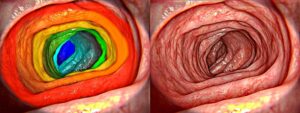
Most endoscopic procedures rely on conventional 2D image sensors for navigation, diagnosis and surgery. In some cases, digital aids such as stereo cameras, endocytoscopy or confocal endoscopy are also implemented. While these modalities add resolution and sometimes give additional depth information, they remain in the world of 2D, failing to penetrate opaque tissue to see what is going on underneath.

Intravenous Ultrasound

To overcome such challenges, special endoscopic ultrasound probes which have proven to be invaluable diagnostic tools, are added to standard procedures; their primary purpose is to detect specific cardiovascular diseases and other problems in blood vessels. They are used, for example, to measure lumen of veins to identify areas of stenosis which could be the result of a birth defect or caused by injury, or to detect lesions which could lead to the narrowing of the arteries or calcification.

Analysis

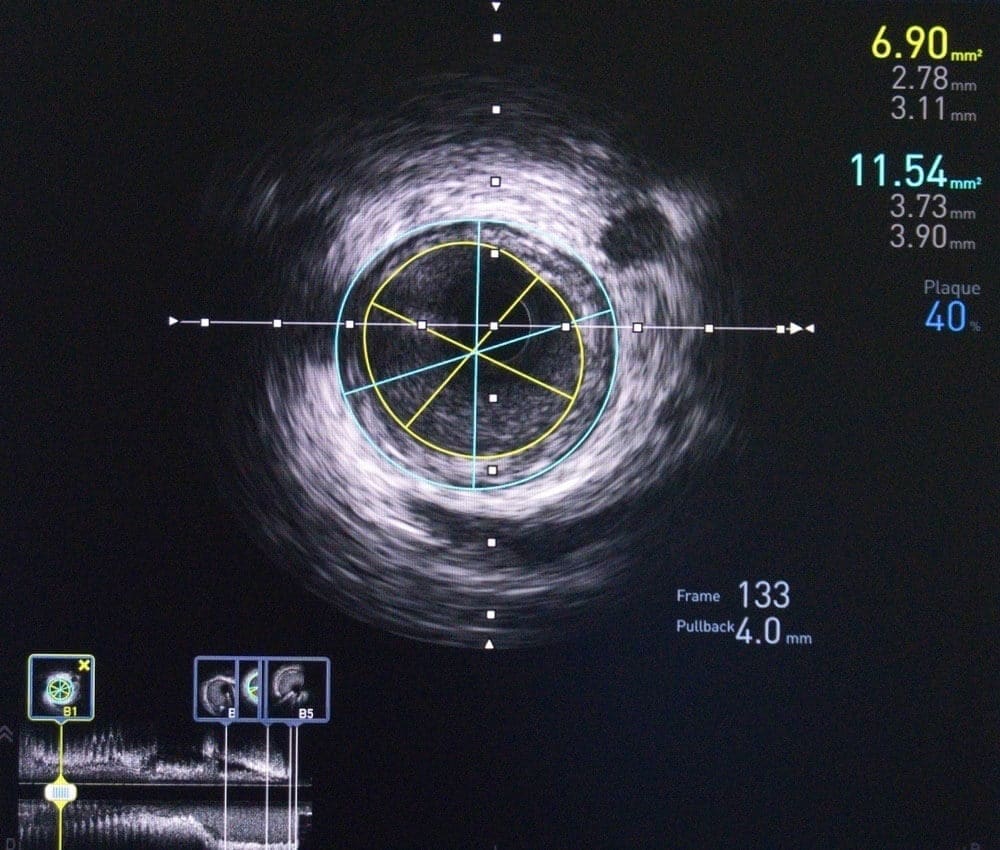
For years, Intravenous Ultrasound (IVUS) has been successfully used to diagnose cardiovascular disease. As the IVUS probe is guided along the blood vessels, at each point a 2D cross section is imaged and eventually a full 3D image of the vessel is created. Next, the image is analyzed in order to measure the lumen and identify areas of stenosis; a process that requires many of hours manual work and work by a radiology specialist. On the other hand, if we employ state of the art image processing techniques, most of the work will be carried-out by a computer, requiring the radiologist to provide only the diagnosis.

AI

Classical computer vision techniques (such as external region analysis or active contour model for example) are able to deal with the relatively easily easy cases, with cross-section that has almost complete cylindrical symmetry. In more challenging cases, deep learning solutions which are based on convolutional neural networks are required to significantly improve results. Deep learning-based approaches have an additional advantage as they can run in real time with the aid of a GPU, and provide immediate feedback to the clinician during the procedure. This allows the clinician to perform a more thorough examination of areas identified to have stenosis, and obtain a more accurate final diagnosis.

RSIP Vision’s CTO Ilya Kovler comments: “From my experience in a multitude of ultrasound projects, I can testify that the speckles noise is a major challenge in any segmentation and measurement task. Using deep learning technology and training with professional cardiologists, you can get excellent results in segmenting the different vessels.”

Share

Share on linkedin
Share on twitter
Share on facebook

Main Field

Endoscopy

AI in Endoscopy is a new field, utilizing innovative AI technologies to enhance the accuracy of Endoscopy imaging and provide better detection and clinical outcomes. RSIP Vision is a pioneer in the field of artificial intelligence for endoscopy imaging. Thanks to our deep learning solutions, radiologists and physicians obtain much more information than before from endoscopy images for both detection of diseases and measurement of therapy progress effectiveness. We have successfully completed a large number of important R&D projects for all kind of minimally invasive procedures. Contact us and discuss your project with our experts!

View Endoscopy

Categories

  • Endoscopy, RSIP Vision Learns, Ultrasound

Related Content

Healthy gut - 3D Rendering

Real time SLAM in Endoscopy Applications

Intravenous OCT

Endoscopic Procedures using Intravenous OCT

Surgical Instrument Segmentation

Surgical Workflow Analysis

AI-based Surgical Workflow Analysis, another big step towards the future of robotic surgery

Healthy gut - 3D Rendering

Real time SLAM in Endoscopy Applications

Intravenous OCT

Endoscopic Procedures using Intravenous OCT

Surgical Instrument Segmentation

Surgical Workflow Analysis

AI-based Surgical Workflow Analysis, another big step towards the future of robotic surgery

Show all

RSIP Vision

Field-tested software solutions and custom R&D, to power your next medical products with innovative AI and image analysis capabilities.

Read more about us

Get in touch

Please fill the following form and our experts will be happy to reply to you soon

Recent News

PR – Intra-op Virtual Measurements in Laparoscopic and Robotic-Assisted Surgeries

PR – Non-Invasive Planning of Coronary Intervention

PR – Bladder Panorama Generator and Sparse Reconstruction Tool

PR – Registration Module for Orthopedic Surgery

All news
Upcoming Events
Stay informed for our next events
Subscribe to Our Magazines

Subscribe now and receive the Computer Vision News Magazine every month to your mailbox

 
Subscribe for free
Follow us
Linkedin Twitter Facebook Youtube

contact@rsipvision.com

Terms of Use

Privacy Policy

© All rights reserved to RSIP Vision 2021

Created by Shmulik

  • Our Work
    • title-1
      • Ophthalmology
      • Uncategorized
      • Ophthalmology
      • Pulmonology
      • Cardiology
      • Orthopedics
    • Title-2
      • Orthopedics
  • Success Stories
  • Insights
  • The company