Skip to content
  • Our Work
    • Fields
      • Cardiology
      • ENT
      • Gastro
      • Orthopedics
      • Ophthalmology
      • Pulmonology
      • Surgical Intelligence
      • Surgical Robotics
      • Urology
      • Other
    • Modalities
      • Endoscopy
      • Medical Segmentation
      • Microscopy
      • Ultrasound
  • Success Stories
  • Insights
    • Magazine
    • Upcoming Events
    • Webinars
    • Meetups
    • News
    • Blog
  • The company
    • About us
    • Careers
Menu
  • Our Work
    • Fields
      • Cardiology
      • ENT
      • Gastro
      • Orthopedics
      • Ophthalmology
      • Pulmonology
      • Surgical Intelligence
      • Surgical Robotics
      • Urology
      • Other
    • Modalities
      • Endoscopy
      • Medical Segmentation
      • Microscopy
      • Ultrasound
  • Success Stories
  • Insights
    • Magazine
    • Upcoming Events
    • Webinars
    • Meetups
    • News
    • Blog
  • The company
    • About us
    • Careers
Contact

Magnetic Resonance Imaging software

Magnetic resonance imaging is a ubiquitous clinical imaging modality, in which patients are placed in a device inducing a magnetic field, and the relaxation of exited hydrogen atoms are collected to form an image of an organ or their entire body. The size of MRI machines makes them practically immobile, whereas the insight gained from their imaging is a desirable properties, which needs to be mobilized and used for finer more precise operations.

Magnetic Resonance Imaging Software - Catheter

This is exactly what our clients were aiming at, when they devised an MRI examination, which can be localized to gain insight into the internal structure of blood vessels. The goals were to obtain a picture from within the vessel, to be able to pinpoint problems of fatty tissues and epithelial lining, and to allow more accurate and safe placement of stents and operations of cardiological surgeries.

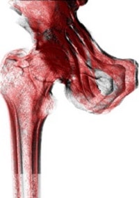
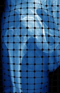
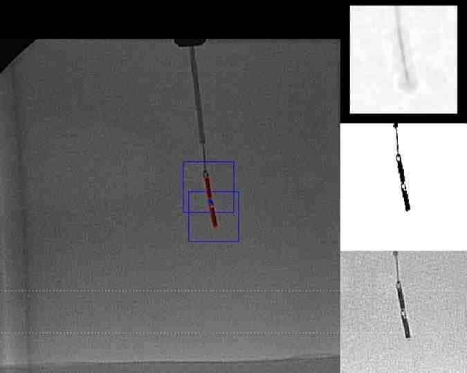
The localized MRI works by inserting rectangular shaped catheter (see image above) through patients’ vascular system and careful navigation up to the target vessel. At the target site, the catheter is carefully rotated on its axis to capture 360 image of the vessel. To construct the MRI image, the orientation of the catheter needs to be correlated to the image obtained at each moment of acquisition, to be able to construct the 3D vessel. However, fine trembling, patient motion and operator faults will result in the catheter not rotating smoothly and at equal speed, and therefore the sequence of acquisitions cannot be reconstructed to give a reliable 3D surface or panoramic image.
 

Magnetic Resonance Imaging software by RSIP Vision


For this end, RSIP Vision has constructed an image processing and computer vision-based algorithm, which can detect the orientation, speed and direction of motion of the rectangular catheter’s heads, as if continuously being moved by the operator. Challenges of low signal to noise ratio were successfully dealt with throughout the project, to accurately extract the orientation of the catheter. We then obtained a one-to-one correspondence between the orientation of the catheters and the acquired image and stitched the images in such a way that they match the correct spatial position of the catheter within the vessel. We thus generated a precise image of the fibrous or fatty tissues in a vessel.

RSIP Vision has been constructing tailor-made algorithmic solutions to the medical industry for over 2 decades. We put emphasis on meeting the accuracy constrains imposed by our clients, allowing them to surpass industrial and regulatory gold standard and push their product to the forefront of technology. To learn more about RSIP Vision’s projects in the medical field, please visit our project pages.

Share

Share on linkedin
Share on twitter
Share on facebook

Main Field

General Medical

View General Medical

Categories

  • General Medical, RSIP Vision Learns

Related Content

Automated RECIST score

Detection and Tracking of Tumors

Pharma - RECIST score

Automated RECIST Measurement

Navigation Systems for Surgery

Catheter Navigation System with RL

U-Net network architecture

Deep Learning in Medical Imaging

Registration of CT and X-ray

Distorted Scan

Calibration of X-ray images

Show all

RSIP Vision

Field-tested software solutions and custom R&D, to power your next medical products with innovative AI and image analysis capabilities.

Read more about us

Get in touch

Please fill the following form and our experts will be happy to reply to you soon

Recent News

PR – Intra-op Virtual Measurements in Laparoscopic and Robotic-Assisted Surgeries

PR – Non-Invasive Planning of Coronary Intervention

PR – Bladder Panorama Generator and Sparse Reconstruction Tool

PR – Registration Module for Orthopedic Surgery

All news
Upcoming Events
Stay informed for our next events
Subscribe to Our Magazines

Subscribe now and receive the Computer Vision News Magazine every month to your mailbox

 
Subscribe for free
Follow us
Linkedin Twitter Facebook Youtube

contact@rsipvision.com

Terms of Use

Privacy Policy

© All rights reserved to RSIP Vision 2021

Created by Shmulik

  • Our Work
    • title-1
      • Ophthalmology
      • Uncategorized
      • Ophthalmology
      • Pulmonology
      • Cardiology
      • Orthopedics
    • Title-2
      • Orthopedics
  • Success Stories
  • Insights
  • The company