Skip to content
  • Our Work
    • Fields
      • Cardiology
      • ENT
      • Gastro
      • Orthopedics
      • Ophthalmology
      • Pulmonology
      • Surgical Intelligence
      • Surgical Robotics
      • Urology
      • Other
    • Modalities
      • Endoscopy
      • Medical Segmentation
      • Microscopy
      • Ultrasound
  • Success Stories
  • Insights
    • Magazine
    • Upcoming Events
    • Webinars
    • Meetups
    • News
    • Blog
  • The company
    • About us
    • Careers
Menu
  • Our Work
    • Fields
      • Cardiology
      • ENT
      • Gastro
      • Orthopedics
      • Ophthalmology
      • Pulmonology
      • Surgical Intelligence
      • Surgical Robotics
      • Urology
      • Other
    • Modalities
      • Endoscopy
      • Medical Segmentation
      • Microscopy
      • Ultrasound
  • Success Stories
  • Insights
    • Magazine
    • Upcoming Events
    • Webinars
    • Meetups
    • News
    • Blog
  • The company
    • About us
    • Careers
Contact

Catheter Navigation System with RL

The typical catheter navigation system relies on fluoroscopy, which exposes patients to dangerous irradiation. To limit the dose throughout the operation, endoscopic cameras are used to guide catheter near the target site of the procedure. When vessel diameter becomes too small to insert an endoscopic camera, magnetic resonance imaging (MRI), CT or ultrasound scans are used to image target site in either the pre-planning stage or at several intervals during operation. This turns navigation into an almost blind intervention and relies on periodic imaging of the area of operation and extraction of quantitative measures to aid surgeons in the guide of catheters. Therefore, it remains a great challenge to plan and navigate catheter into soft tissue, to be able to operate on site, to sample tissue for biopsy, to perform ablation and so on, with minimal harm to patient and through the shortest possible path of catheter insertion.

Navigation Systems for Surgery

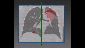
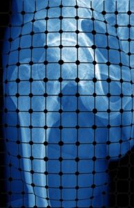
To construct a vision-aided catheter navigation system, the positions of blood vessels and segmented airways along the planned path of the catheter need to be accounted for. This allows to generate the shortest and safest catheter insertion trajectory, while allowing to plan the intentional puncture of non-critical tissues using the catheter, in order to arrive at a target site. Because CT scans are taken at static patient position, the real time imaging (fluorophore) needs to be registered with the airways and the vascular tree, and catheter position tracked over time. The irradiation dose needs to be reduced to non-damaging levels, resulting in poorly contrasted and noisy images of soft tissues. Recent developments in catheter hardware technology provide the means of acquiring additional local information based on MRI within vessels. Furthermore, sensors placed on catheter tips allow to detect whether the catheter is near lumen or vessel walls. This information can be integrated together to form a much clearer picture, while working almost blindly in a vessel which no endoscopic camera can have access to.

Path planning using the integrated sources of information (CT, airways segmentation and the like) can be done in several settings. A straightforward approach would be to utilize graph path techniques (e.g., Dijkstra, Bellman-Ford, etc.), where edges and nodes of the graph are defined by the architecture of the segmented vascular tree and the cost of the path is determined by measures of safety and other patient-specific physical constraints. This approach by itself, cannot account for off-vessel paths and therefore it needs to be reinforced by additional edges, which represent punctured paths. A complication arises when such paths are non-linear, and a tunnel needs to be dug inside a tissue. Hence, for this non-linear constraint it is most productive to work in the framework of machine learning.

Reinforcement Learning to support catheter navigation systems

Reinforcement Learning methods can be used to simulate the path taken by experts in a test setting, where negative rewards are given for dangerous paths and tunnels punctured. A combination of the graph optimization techniques, combined with Reinforcement learning for off-vessel planning, can provide the safest catheter trajectory path towards target. This and other highly complex algorithms are being constructed daily by the engineers of RSIP Vision. Our company is a global leader in constructing tailor-made computer vision, image processing and machine learning solutions for medical and other industrial domains. We have successfully done this for many years. Please use the contact page to consult our experts about your project. To learn more about RSIP Vision’s solutions and fields of expertise, please visit our projects page.

Share

Share on linkedin
Share on twitter
Share on facebook

Main Field

General Medical

View General Medical

Categories

  • General Medical, RSIP Vision Learns

Related Content

Automated RECIST score

Detection and Tracking of Tumors

Pharma - RECIST score

Automated RECIST Measurement

U-Net network architecture

Deep Learning in Medical Imaging

Magnetic Resonance Imaging Software - Catheter

Magnetic Resonance Imaging software

Registration of CT and X-ray

Distorted Scan

Calibration of X-ray images

Show all

RSIP Vision

Field-tested software solutions and custom R&D, to power your next medical products with innovative AI and image analysis capabilities.

Read more about us

Get in touch

Please fill the following form and our experts will be happy to reply to you soon

Recent News

PR – Intra-op Virtual Measurements in Laparoscopic and Robotic-Assisted Surgeries

PR – Non-Invasive Planning of Coronary Intervention

PR – Bladder Panorama Generator and Sparse Reconstruction Tool

PR – Registration Module for Orthopedic Surgery

All news
Upcoming Events
Stay informed for our next events
Subscribe to Our Magazines

Subscribe now and receive the Computer Vision News Magazine every month to your mailbox

 
Subscribe for free
Follow us
Linkedin Twitter Facebook Youtube

contact@rsipvision.com

Terms of Use

Privacy Policy

© All rights reserved to RSIP Vision 2021

Created by Shmulik

  • Our Work
    • title-1
      • Ophthalmology
      • Uncategorized
      • Ophthalmology
      • Pulmonology
      • Cardiology
      • Orthopedics
    • Title-2
      • Orthopedics
  • Success Stories
  • Insights
  • The company