Reconstruction of the 3D shape of a surface viewed under the microscope poses multiple challenges. The surfaces might take on highly irregular shapes in the resolution viewed, hence making reconstruction by light reflection (shape from shading) highly unstable and inaccurate.
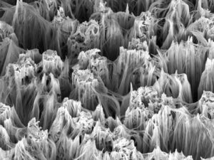
Irregular, non-smooth, surfaces having many sharp bends and peaks, can be characterized as having a high frequency texture pattern. Quite often, high frequency information need to be ignored or smoothed out, in order to be able to stably reconstruct a surface. Essentially, what we search for is a low-pass filter for those images, meaning a circuit offering an easy passage to low-frequency signals and a difficult passage to high-frequency signals. Simple kernels like the Gaussian can take the role of such low-pass filter, paving the way to scale-space methods to detect salient features in the object under the microscope. However, for reconstruction purposes, we need to evaluate the object’s depth by varying the focal plane.
The shape-from-focus method, developed about two decades ago, provides us with the framework to do just that. In shape-from-focus, a sequence of images is acquired where each image has a different focus level. The relative degree of focus between the images is evaluated by using the sum-modified Gaussian transformation. This transformation produces in each pixel a sum of Laplacian operators on the pixel values and its neighbors’. A thresholding, or maxima operator, is then applied to the transformed image stack, to determine the depth of the surface in each pixel.
Reconstruction of microscopic surface, specifically the rough ones, has been demonstrated to perform well using shape from focus. The shape from focus methodology provides a simple framework to assess the geometrical properties of an object, even in different illumination and reflectance conditions. The advantages of shape from focus stems from its ability to treat high frequency surface, like the membrane of a cell (or that of a virus) seen in electron microscopy, to reveal novel structures and interaction dependent on volumetric information.
As a computer vision specialist, RSIP Vision has a vast experience in the reconstruction of structures for the use of biomedical and semiconductor industries. Contact us to learn more about the in-house expertise accrued by RSIP Vision, which allows us to deliver cutting-edge solutions for the reconstruction of both anatomical and microscopic objects.
 Microscopy