Skip to content
  • Our Work
    • Fields
      • Cardiology
      • ENT
      • Gastro
      • Orthopedics
      • Ophthalmology
      • Pulmonology
      • Surgical Intelligence
      • Surgical Robotics
      • Urology
      • Other
    • Modalities
      • Endoscopy
      • Medical Segmentation
      • Microscopy
      • Ultrasound
  • Success Stories
  • Insights
    • Magazine
    • Upcoming Events
    • Webinars
    • Meetups
    • News
    • Blog
  • The company
    • About us
    • Careers
Menu
  • Our Work
    • Fields
      • Cardiology
      • ENT
      • Gastro
      • Orthopedics
      • Ophthalmology
      • Pulmonology
      • Surgical Intelligence
      • Surgical Robotics
      • Urology
      • Other
    • Modalities
      • Endoscopy
      • Medical Segmentation
      • Microscopy
      • Ultrasound
  • Success Stories
  • Insights
    • Magazine
    • Upcoming Events
    • Webinars
    • Meetups
    • News
    • Blog
  • The company
    • About us
    • Careers
Contact

Trajectory tracking of a fluorescent tag

Limitation of microscopic modalities to directly observe sub-cellular structures has led to the development of indirect measuring procedures based on single particle tracking. Analysis of trajectories of a fluorescently tagged particle allows indirect estimation of the spatial organization of the domain confining its motion.

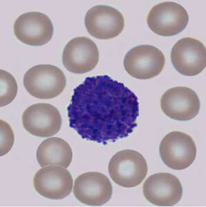
Single particle trajectories of several tagged monomers (molecular blocks forming more complex molecules, called polymers) can reveal the interaction between organelles (tiny cellular organs, suspended in the cytoplasm of a eukaryotic cell) like telomeres (the extremities of a chromosome, which protect it from deterioration) with the nuclear membrane or among themselves. Quantitative temporal information, such as the mean first arrival time of tagged particle to a region of interest can be extracted. One such example is the study of the average time it takes a virus to travel from the cell membrane to a nuclear pore.

Learning the behavior of bio-molecules and the interaction they have with other molecular structures in their native environment gives us valuable information about processes like viral infections of cells, protein-DNA interactions and other complex biological processes.

Microscopy imaging of metastatic cancer cells

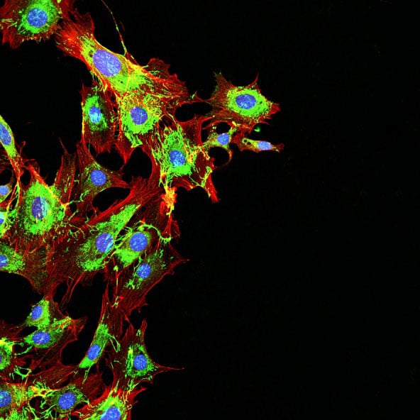
Tracking of a fluorescent tag can be initialized by either manually marking the point of interest, or automatically detecting all tags in the first frame. The difficulty in detecting and tracking particles in dynamic live microscopy of fluorescent tags resides mainly in the high levels of noise. Increasing the signal-to-noise ratio of tags in images is achieved by filtering the image with Gaussian or Laplacian-like kernels. The use of these kernels is motivated by the Gaussian intensity signature of the tagged particle.

Localization of the tagged particle in each frame is performed after stabilization and alignment of the image sequence. This step is required to compensate for small sample motion throughout the acquisition period. In a sequence of segmented tag positions, tracking is regarded as the process of connecting the dots between corresponding tags of subsequent frames.

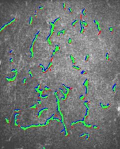
The task is to find an optimal path in time representing the trajectory. The problem can be regarded as a minimization of a cost function, having penalizing term on sharp jumps in particle displacement. In addition, image intensity levels are integrated into the cost function to favor bright spots. The minimization problem can be solved in the framework of dynamic programming.

The result is a path with minimal cost, representing three-dimensional coordinates of a fluorescent tag particle throughout the image sequence. Tracking objects in a sequence of images is one of RSIP Vision’s fields of expertise. We have been utilizing dynamic programming and graph theoretic approaches to track not only tags, but also full organs, like in this heart monitoring system project.
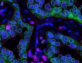
Share

Share on linkedin
Share on twitter
Share on facebook

Main Field

Microscopy

View Microscopy

Categories

  • Microscopy, RSIP Vision Learns

Related Content

Multiplex

Multiplex IF Analysis

CTCs - Circulating Tumor Cells

Circulating Tumor Cells (CTCs)

Pharma - Tissue Analysis

Tissue Analysis with AI

Pharma - Dendritic cell

Detection and Segmentation of Dendritic Cells

Cell Classification - pattern recognition classification techniques

Biomedical image processing and algorithms

Microscopy view of Monocytes

Cell Classification software

Show all

RSIP Vision

Field-tested software solutions and custom R&D, to power your next medical products with innovative AI and image analysis capabilities.

Read more about us

Get in touch

Please fill the following form and our experts will be happy to reply to you soon

Recent News

PR – Intra-op Virtual Measurements in Laparoscopic and Robotic-Assisted Surgeries

PR – Non-Invasive Planning of Coronary Intervention

PR – Bladder Panorama Generator and Sparse Reconstruction Tool

PR – Registration Module for Orthopedic Surgery

All news
Upcoming Events
Stay informed for our next events
Subscribe to Our Magazines

Subscribe now and receive the Computer Vision News Magazine every month to your mailbox

 
Subscribe for free
Follow us
Linkedin Twitter Facebook Youtube

contact@rsipvision.com

Terms of Use

Privacy Policy

© All rights reserved to RSIP Vision 2021

Created by Shmulik

  • Our Work
    • title-1
      • Ophthalmology
      • Uncategorized
      • Ophthalmology
      • Pulmonology
      • Cardiology
      • Orthopedics
    • Title-2
      • Orthopedics
  • Success Stories
  • Insights
  • The company