Skip to content
  • Our Work
    • Fields
      • Cardiology
      • ENT
      • Gastro
      • Orthopedics
      • Ophthalmology
      • Pulmonology
      • Surgical Intelligence
      • Surgical Robotics
      • Urology
      • Other
    • Modalities
      • Endoscopy
      • Medical Segmentation
      • Microscopy
      • Ultrasound
  • Success Stories
  • Insights
    • Magazine
    • Upcoming Events
    • Webinars
    • Meetups
    • News
    • Blog
  • The company
    • About us
    • Careers
Menu
  • Our Work
    • Fields
      • Cardiology
      • ENT
      • Gastro
      • Orthopedics
      • Ophthalmology
      • Pulmonology
      • Surgical Intelligence
      • Surgical Robotics
      • Urology
      • Other
    • Modalities
      • Endoscopy
      • Medical Segmentation
      • Microscopy
      • Ultrasound
  • Success Stories
  • Insights
    • Magazine
    • Upcoming Events
    • Webinars
    • Meetups
    • News
    • Blog
  • The company
    • About us
    • Careers
Contact

3D Cardiac MRI automatic segmentation

Massive progress in 3D Cardiac magnetic resonance imaging (MRI) gives today access to good 3D mapping of the heart, using gating to synchronize to the systolic phase of the pulse. MRI can give good distinction between the different tissues and blood without any hazard connected to radiation.


Cardiac MRI 3D automatic segmentation

3D Cardiac MRI has an important role in diagnosis, pre-operative planning, and post-operative management of patients with cardiac pathologies. Fully automated segmentation of these images will enable faster and more reliable extraction of data from these images, helping practitioners take more accurate treatment decisions.

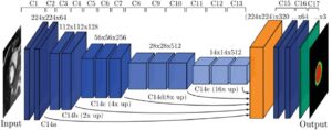
In our project, based on state-of-the-art deep learning techniques, we demonstrate multiclass segmentation of 3D cardiac MRI using a fully convolutional neural network (CNN) with a Unet-based architecture.

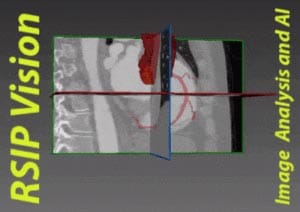
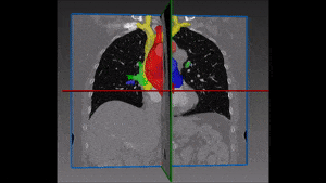
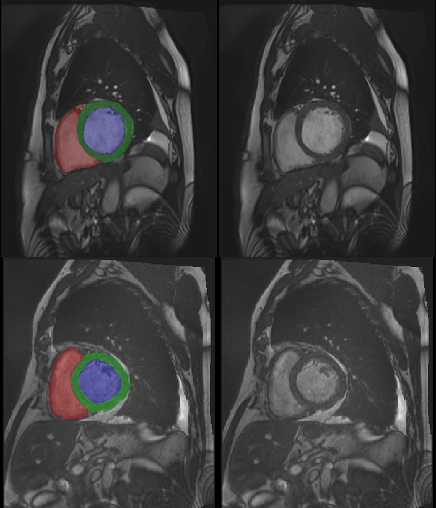
The deep learning network performs automatic segmentation of the right ventricle (red in the image above) left ventricle (blue) and myocardium (green), for both end diastole and end systole phases.

Challenges in 3D Cardiac MRI automatic segmentation


The main challenges of this algorithmic procedure are as follows:
3D data: dealing with 3D data makes for a very large network due to the need for 3D convolution kernels. Since available databases are not very large, this is a major challenge. To overcome it, we have found the correct balance for the size of patches generated from the dataset. They should be small enough to allow sufficient training examples to be generated; but also large enough to contain sufficient information about the region of interest.

Initialization: the size of the CNN also makes it very sensitive to the type of initialization which is used on the convolution kernels. This factor alone can determine if the network will converge or not. We found that the best method in this case was uniform initialization.

CNN for Cardiac MRI

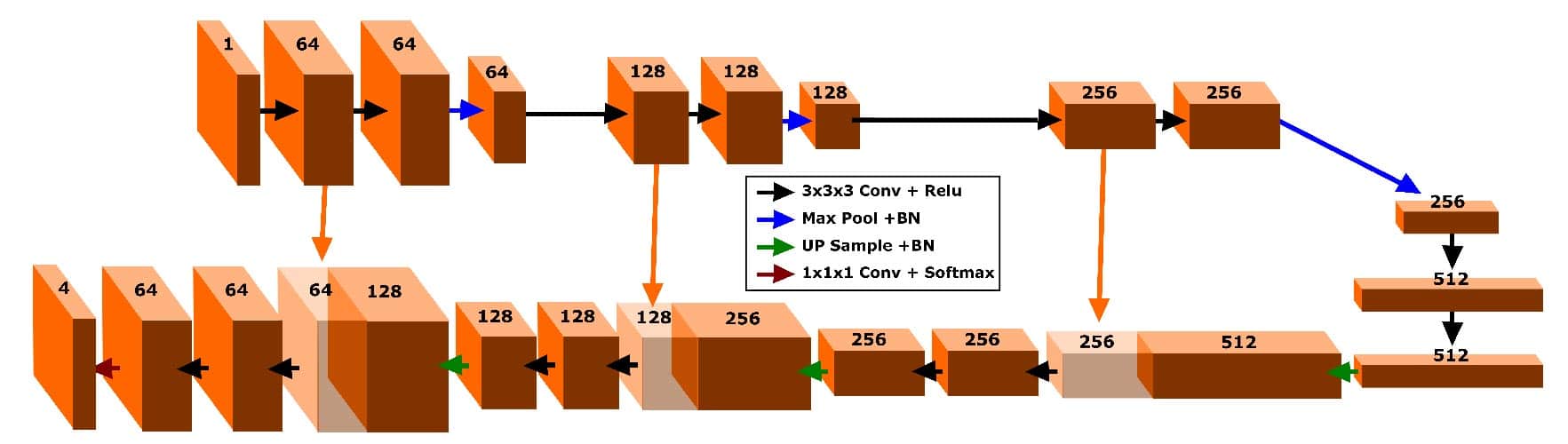
Memory – Left and right cardiac ventricles are large, which means that in order to detect them accurately a large input is needed. When GPU memory is limited, it is necessary to find the optimal tradeoff between input size and batch size. One wants to use as large a batch as possible (for normalization reasons), but on the other hand, a large input which can consume vast amounts of memory.

3D cardiac MRI with DL

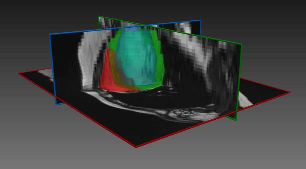
Tackling these challenges, segmentation technology with deep learning by RSIP Vision achieves fast, robust and accurate segmentation of the right ventricle (red) left ventricle (blue) and myocardium (green), for both end diastole and end systole phases. The multiple classes are extracted simultaneously, in constant running time, while the software is easily deployable on standard PC hardware with standard NVidia GPU. Our approach for Artificial Intelligence in cardiology proves to be very effective. The technology presented above for 3D Cardiac MRI automatic segmentation can also be adapted to segment other organs under other imaging modalities, given appropriate training dataset.

Would you like to know how RSIP Vision’s technology and experience can help you obtain better results from your network? Contact our experts now!

Share

Share on linkedin
Share on twitter
Share on facebook

Main Field

Cardiology

RSIP Vision’s advanced algorithms and image processing software (together with proper machines and devices) are used by physicians and researchers to develop and perform preventive and interventional cardiovascular procedures: that's our contribution to Artificial Intelligence in cardiology. Our software provides information about the patient’s cardiovascular system in a non-invasive manner, helps physicians ensure accuracy during complex operations, raises the level of patient safety through limiting his/her exposure to radiation, and aids diagnosis. We come to the office to write algorithms that save lives. In this section about computer vision in cardiology you can discover some of the ways we do it.

View Cardiology

Categories

  • Cardiology, RSIP Vision Learns

Related Content

PR – Non-Invasive Planning of Coronary Intervention

AI in Cardiac CT angiography

Coronary Arteries Segmentation

Great Vessels Segmentation with Deep Learning

Cardiac Motion Correction

Deep Learning in Cardiology

3D Reconstruction of the heart

3D reconstruction of the heart

PR – Non-Invasive Planning of Coronary Intervention

AI in Cardiac CT angiography

Coronary Arteries Segmentation

Great Vessels Segmentation with Deep Learning

Cardiac Motion Correction

Deep Learning in Cardiology

3D Reconstruction of the heart

3D reconstruction of the heart

Show all

RSIP Vision

Field-tested software solutions and custom R&D, to power your next medical products with innovative AI and image analysis capabilities.

Read more about us

Get in touch

Please fill the following form and our experts will be happy to reply to you soon

Recent News

PR – Intra-op Virtual Measurements in Laparoscopic and Robotic-Assisted Surgeries

PR – Non-Invasive Planning of Coronary Intervention

PR – Bladder Panorama Generator and Sparse Reconstruction Tool

PR – Registration Module for Orthopedic Surgery

All news
Upcoming Events
Stay informed for our next events
Subscribe to Our Magazines

Subscribe now and receive the Computer Vision News Magazine every month to your mailbox

 
Subscribe for free
Follow us
Linkedin Twitter Facebook Youtube

contact@rsipvision.com

Terms of Use

Privacy Policy

© All rights reserved to RSIP Vision 2021

Created by Shmulik

  • Our Work
    • title-1
      • Ophthalmology
      • Uncategorized
      • Ophthalmology
      • Pulmonology
      • Cardiology
      • Orthopedics
    • Title-2
      • Orthopedics
  • Success Stories
  • Insights
  • The company