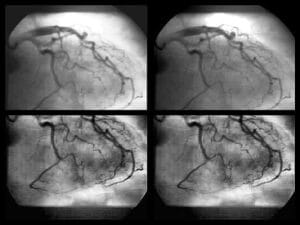
Coronary computed tomography angiography (CCTA) is an efficient and non-invasive imaging modality with widespread clinical implementation in the identification of coronary artery disease (CAD). With rapid 3D visualization of coronary arteries and heart, including visualization of blood flow in arteries and capillaries, CCTA enables accurate monitoring of the coronary tree for blockages and other pathologies, yet, some major challenges remain to be met to maximize its clinical value.
Complexity of CCTA Analysis:
diagnostic accuracy of CCTA.
Deep learning is automating segmentation and facilitating the identification of blockages and other anomalies while increasing accuracy and time savings.
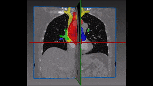
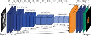
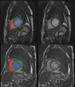
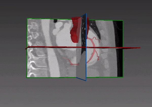
Methods – One DL method is segmenting the LV in CCTA using a multiscale patch-based CNN. This is a two-stage method. In the first stage, three CNNs are dispatched to detect a bounding box around the LV. In the second stage, CNNs are used to perform voxel classification.
Another method involves using a LSTM RNN to automatically label segments in the coronary artery tree, trained using hundreds of CCTA images. The RNN, is proven to successfully learn the topology and spatial location of blood vessels and capture the coronary tree structure characteristics while presenting robust results.
Results – DL based approaches, such as CNN and RNN, successfully segment the coronary tree structure and characteristics, reducing complexity and time burden of CCTA interpretation and achieving a higher accuracy rate than classical approaches. A future challenge is to handle even the most complicated pathologies while maintaining a high accuracy rate.
Stenosis Detection:
CTTA has a high negative predictive value, making this modality a suitable tool for excluding significant coronary artery calcification (CAC). However, despite this high sensitivity, CTTA has limited specificity for identifying atherosclerotic plaque and functionally significant stenosis. Non-conclusive analysis leads to increased rates of invasive procedures, such as fractional flow reserve (FFR) measurement during ICA, and delayed CAD management.
Methods – A two-stage DL method may be used to identify CCTA image voxels that represent coronary artery calcification. The method uses two separate CNNs. The first segments the image and identifies potential calcification, and the second CNN classifies only the voxels selected by the first CNN to identify functional significance. This approach achieves a high rate of accuracy.
Another method uses a Support Vector Machine (SVP) trained to evaluate CCTA images for stenosis, thereby facilitating the identification of patients with functionally significant stenosis.
Results – DL architectures are more sensitive and quicker in identifying functionally significant stenosis than standard CCTA which despite high negative predictive value in exclusion is less efficient at detecting and identifying CAC and functionally significant stenosis.
Low-Dose Contrast Analysis:
The usage of contrast agents in CCTA improves image quality and segmentation accuracy contributing to overall diagnostic efficiency. However, patient safety is compromised by increased dosage.
On the other hand, dose reduction increases noise and artifact interference, impacting image interpretation and making classic CV tolls less effective. DL algorithms are enhancing low dose CCTA image quality, thereby lowering dose radiation for contrast CCTA and improving clinical analysis.
Methods – Different approaches are addressing the above challenges. One uses an FCNN trained on 3D CCTA data patches to predict routine dose images from low dose CCTA data patches. Simulated and real low dose scans were associated with a 90% dose reduction. An additional 2D CNN was utilized for improving denoising.
Other methods use a GAN to denoise low-dose CCTA images and reconstruct low-dose images into routine-dose CCTA images, or use an SSDA-based CNN to reconstruct, denoise, and inpaint low-dose CCTA images.
Results – Several CNNs are effective at predicting and reconstructing low dose CCTA images to emulate the image quality of routine dose CCTA images. These results have been validated, showing that using fast and high-quality DL algorithmic denoising may contribute to radiation dose reduction of 90%, thereby improving patient safety while enhancing image quality.
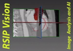
RSIP at the Heart of Cardiac Imaging
RSIP Vision is a first-mover in deep learning for CT, at the heart of AI for cardiac care. Click here for more information about our computer vision solutions for cardiology.
 Cardiology