Skip to content
  • Our Work
    • Fields
      • Cardiology
      • ENT
      • Gastro
      • Orthopedics
      • Ophthalmology
      • Pulmonology
      • Surgical Intelligence
      • Surgical Robotics
      • Urology
      • Other
    • Modalities
      • Endoscopy
      • Medical Segmentation
      • Microscopy
      • Ultrasound
  • Success Stories
  • Insights
    • Magazine
    • Upcoming Events
    • Webinars
    • Meetups
    • News
    • Blog
  • The company
    • About us
    • Careers
Menu
  • Our Work
    • Fields
      • Cardiology
      • ENT
      • Gastro
      • Orthopedics
      • Ophthalmology
      • Pulmonology
      • Surgical Intelligence
      • Surgical Robotics
      • Urology
      • Other
    • Modalities
      • Endoscopy
      • Medical Segmentation
      • Microscopy
      • Ultrasound
  • Success Stories
  • Insights
    • Magazine
    • Upcoming Events
    • Webinars
    • Meetups
    • News
    • Blog
  • The company
    • About us
    • Careers
Contact

Great Vessels Segmentation with Deep Learning

The great vessels conduct blood to and from the heart. These vessels include the aorta, superior and inferior vena cava, pulmonary arteries and pulmonary veins. Since the great vessels are an integral part of systemic and pulmonary circulation, vascular pathologies involving said vessels might be deadly.

Great Vessels Segmentation
Great Vessels segmentation: red = aorta; yellow = superior vena cava; blue = pulmonary artery; green = pulmonary veins

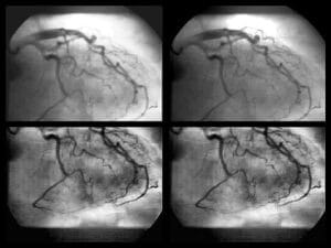
Computed tomography (CT) imaging is a useful tool for assessing the great vessels, and has become an essential tool for diagnosis of vascular pathologies, including aortic arch aneurysm, valvular heart disease and congenital vascular anomalies, such as aortic stenosis, coarctation of aorta or transposition of great vessels.

Furthermore, accurate imaging helps with surgical planning or endovascular procedures e.g. endovascular aneurysm repair (EVAR) or transcatheter aortic valve implantation (TAVI). Furthermore, it might even aid with quick diagnosis and surgical planning during life threatening vascular medical emergencies, such as aortic dissection or ruptured aortic aneurysm.

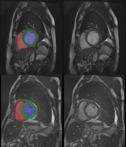
Moreover, by identifying vessels within the mediastinum, the physician can easily distinguish between vessels and other important structures including the heart, esophagus and trachea, or even abnormal masses. Hence, 3D segmentation of the great vessels proves to be extremely helpful as a diagnostic tool, surgical planning and guidance tool, as well as for follow-up.

Automated Great Vessels Segmentation:

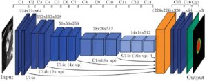
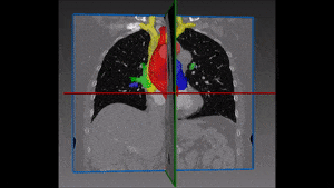
Utilizing deep convolutional networks, RSIP Vision’s engineers built a cutting-edge AI module for automatic segmentation of the great vessels. Combining global context and local information from the mediastinum area, the module is able to overcome the challenge posed by the target’s high anatomical variability. RSIP Vision’s fast and accurate module does not require any human intervention – making it the ideal instrument for identification and segmentation of great vessels in CT scans. Call us to discuss your cardio segmentation plans and take us along for your Deep Learning projects!
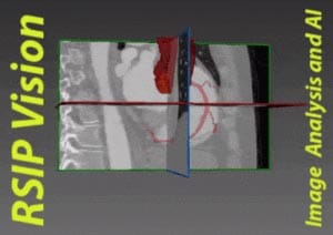
Share

Share on linkedin
Share on twitter
Share on facebook

Main Field

Cardiology

RSIP Vision’s advanced algorithms and image processing software (together with proper machines and devices) are used by physicians and researchers to develop and perform preventive and interventional cardiovascular procedures: that's our contribution to Artificial Intelligence in cardiology. Our software provides information about the patient’s cardiovascular system in a non-invasive manner, helps physicians ensure accuracy during complex operations, raises the level of patient safety through limiting his/her exposure to radiation, and aids diagnosis. We come to the office to write algorithms that save lives. In this section about computer vision in cardiology you can discover some of the ways we do it.

View Cardiology

Categories

  • Cardiology, RSIP Vision Learns

Related Content

PR – Non-Invasive Planning of Coronary Intervention

AI in Cardiac CT angiography

Coronary Arteries Segmentation

Cardiac Motion Correction

Deep Learning in Cardiology

Cardiac MRI 3D automatic segmentation

3D Cardiac MRI automatic segmentation

3D Reconstruction of the heart

3D reconstruction of the heart

Show all

RSIP Vision

Field-tested software solutions and custom R&D, to power your next medical products with innovative AI and image analysis capabilities.

Read more about us

Get in touch

Please fill the following form and our experts will be happy to reply to you soon

Recent News

PR – Intra-op Virtual Measurements in Laparoscopic and Robotic-Assisted Surgeries

PR – Non-Invasive Planning of Coronary Intervention

PR – Bladder Panorama Generator and Sparse Reconstruction Tool

PR – Registration Module for Orthopedic Surgery

All news
Upcoming Events
Stay informed for our next events
Subscribe to Our Magazines

Subscribe now and receive the Computer Vision News Magazine every month to your mailbox

 
Subscribe for free
Follow us
Linkedin Twitter Facebook Youtube

contact@rsipvision.com

Terms of Use

Privacy Policy

© All rights reserved to RSIP Vision 2021

Created by Shmulik

  • Our Work
    • title-1
      • Ophthalmology
      • Uncategorized
      • Ophthalmology
      • Pulmonology
      • Cardiology
      • Orthopedics
    • Title-2
      • Orthopedics
  • Success Stories
  • Insights
  • The company