Skip to content
  • Our Work
    • Fields
      • Cardiology
      • ENT
      • Gastro
      • Orthopedics
      • Ophthalmology
      • Pulmonology
      • Surgical Intelligence
      • Surgical Robotics
      • Urology
      • Other
    • Modalities
      • Endoscopy
      • Medical Segmentation
      • Microscopy
      • Ultrasound
  • Success Stories
  • Insights
    • Magazine
    • Upcoming Events
    • Webinars
    • Meetups
    • News
    • Blog
  • The company
    • About us
    • Careers
Menu
  • Our Work
    • Fields
      • Cardiology
      • ENT
      • Gastro
      • Orthopedics
      • Ophthalmology
      • Pulmonology
      • Surgical Intelligence
      • Surgical Robotics
      • Urology
      • Other
    • Modalities
      • Endoscopy
      • Medical Segmentation
      • Microscopy
      • Ultrasound
  • Success Stories
  • Insights
    • Magazine
    • Upcoming Events
    • Webinars
    • Meetups
    • News
    • Blog
  • The company
    • About us
    • Careers
Contact

Automatic Catheter Orientation Measurement

Measuring Catheter Orientation in MRI

Lipid-rich coronary lesions (atherosclerosis) have a high risk of rupturing and causing severe coronary damage. These lesions are often treated using interventional coronary catheterization for high-risk patients. Early detection and quantification is necessary in order to increase the chances of a successful treatment and prevention.

Magnetic Resonance Imaging Software - Catheter

During the catheterization process, catheters are inserted with measurement equipment at their tips. These sensors scan their immediate surroundings and indicate levels of lipidity and calcium enrichment.

For example, Top Spin Medical developed a unique catheter with MRI-like capabilities, which has been clinically tested and installed in hospitals. Following catheter insertion, the surrounding tissues send pulses to the tip of the catheter that allow measurements of tissue parameters such as its lipid layer or calcium composition. These signals alert the physician regarding the location of any possible lesion. The exact position of the lesion must be pinpointed for the accuracy of the interventional procedure and to decrease the risk during operation. For these reasons, it is crucial to accurately measure the exact orientation the of catheter.

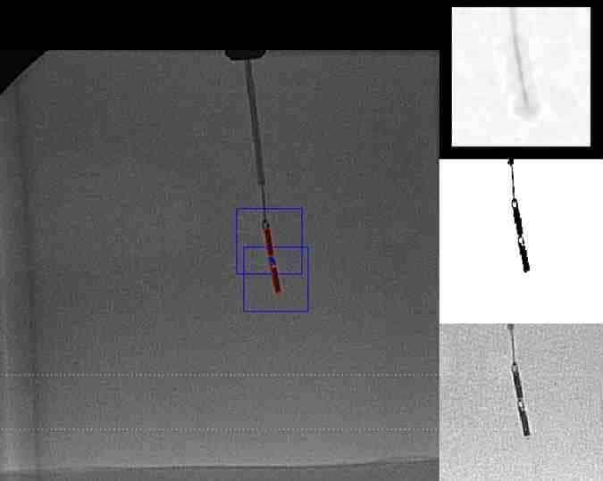
The main problem is that the orientation of the catheter’s tip is unknown throughout the insertion process and so the exact location cannot be accurately given. To view the catheter during insertion and positioning, physicians use local X-ray imaging. In these images the outer edges of the tip are clearly visible. However, X-ray images are two-dimensional, and so when the catheter tip is oriented perpendicular to the plane of the image, the physician cannot tell whether it is pointing inwards or outwards.

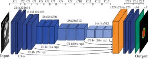
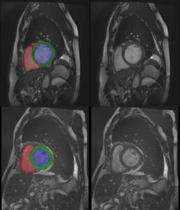
By employing advanced image analysis and deep learning methods, RSIP Vision has constructed a method to ascertain the exact orientation of the catheter tip from X-ray images. Single 2D X-ray images obtained during catheter insertion provide the input to our algorithms. During the first stage, the catheter tip is segmented in the given images. The opacity of the tip in the image is then used as a means of telling its orientation. Using RSIP Vision’s algorithms, not only can the orientation information be used by the physician during catheterization, but additionally more accurate positions of lipid lesions can be given at the examination site.

Share

Share on linkedin
Share on twitter
Share on facebook

Main Field

Cardiology

RSIP Vision’s advanced algorithms and image processing software (together with proper machines and devices) are used by physicians and researchers to develop and perform preventive and interventional cardiovascular procedures: that's our contribution to Artificial Intelligence in cardiology. Our software provides information about the patient’s cardiovascular system in a non-invasive manner, helps physicians ensure accuracy during complex operations, raises the level of patient safety through limiting his/her exposure to radiation, and aids diagnosis. We come to the office to write algorithms that save lives. In this section about computer vision in cardiology you can discover some of the ways we do it.

View Cardiology

Categories

  • Cardiology, RSIP Vision Learns

Related Content

PR – Non-Invasive Planning of Coronary Intervention

AI in Cardiac CT angiography

Coronary Arteries Segmentation

Great Vessels Segmentation with Deep Learning

Cardiac Motion Correction

Deep Learning in Cardiology

Cardiac MRI 3D automatic segmentation

3D Cardiac MRI automatic segmentation

PR – Non-Invasive Planning of Coronary Intervention

AI in Cardiac CT angiography

Coronary Arteries Segmentation

Great Vessels Segmentation with Deep Learning

Cardiac Motion Correction

Deep Learning in Cardiology

Cardiac MRI 3D automatic segmentation

3D Cardiac MRI automatic segmentation

Show all

RSIP Vision

Field-tested software solutions and custom R&D, to power your next medical products with innovative AI and image analysis capabilities.

Read more about us

Get in touch

Please fill the following form and our experts will be happy to reply to you soon

Recent News

PR – Intra-op Virtual Measurements in Laparoscopic and Robotic-Assisted Surgeries

PR – Non-Invasive Planning of Coronary Intervention

PR – Bladder Panorama Generator and Sparse Reconstruction Tool

PR – Registration Module for Orthopedic Surgery

All news
Upcoming Events
Stay informed for our next events
Subscribe to Our Magazines

Subscribe now and receive the Computer Vision News Magazine every month to your mailbox

 
Subscribe for free
Follow us
Linkedin Twitter Facebook Youtube

contact@rsipvision.com

Terms of Use

Privacy Policy

© All rights reserved to RSIP Vision 2021

Created by Shmulik

  • Our Work
    • title-1
      • Ophthalmology
      • Uncategorized
      • Ophthalmology
      • Pulmonology
      • Cardiology
      • Orthopedics
    • Title-2
      • Orthopedics
  • Success Stories
  • Insights
  • The company