Skip to content
  • Our Work
    • Fields
      • Cardiology
      • ENT
      • Gastro
      • Orthopedics
      • Ophthalmology
      • Pulmonology
      • Surgical Intelligence
      • Surgical Robotics
      • Urology
      • Other
    • Modalities
      • Endoscopy
      • Medical Segmentation
      • Microscopy
      • Ultrasound
  • Success Stories
  • Insights
    • Magazine
    • Upcoming Events
    • Webinars
    • Meetups
    • News
    • Blog
  • The company
    • About us
    • Careers
Menu
  • Our Work
    • Fields
      • Cardiology
      • ENT
      • Gastro
      • Orthopedics
      • Ophthalmology
      • Pulmonology
      • Surgical Intelligence
      • Surgical Robotics
      • Urology
      • Other
    • Modalities
      • Endoscopy
      • Medical Segmentation
      • Microscopy
      • Ultrasound
  • Success Stories
  • Insights
    • Magazine
    • Upcoming Events
    • Webinars
    • Meetups
    • News
    • Blog
  • The company
    • About us
    • Careers
Contact

Classification and Segmentation of Dendritic Cells

Dry eye disease (DED) is one of the most common ophthalmic disorders. Inflammation of the ocular surface is controlled by corneal antigen-presenting cells called dendritic cells (DCs), which induce T-cell activation, and play a critical role in the pathogenesis of dry eye disease. The density of corneal DC is correlated with both symptoms and clinical signs in dry eye, making it an attractive non-invasive and responsive surrogate biomarker to assess the severity of corneal inflammation. To evaluate the effect of inflammation on the cornea structures and function, coronal images at a cellular level are acquired in a non-invasive manner by in vivo confocal microscopy (IVCM), to visualize the density of immune and inflammatory cells. Manual classification immune cells in IVCM images is highly time consuming and prone to subjective decisions and levels of expertise: it therefore becomes a bottleneck in the detection and assessment of DED prognosis. However, an automated convolutional neural networks (CNN) for the detection and quantification of dendritic cells in IVCM images can be used as an objective evaluation of ocular surface inflammation to improve both diagnostic and treatment accuracy.
.
 .
For this classification and segmentation of dendritic cells, RSIP vision in collaboration with expert physicians have trained a U-Net based CNN to classify corneal layers, segment and quantify DCs density in four tissue types acquired by IVCM images: epithelium, subbasal nerve plexus (SBP), stroma, and endothelium. U-Net is a fully CNN architecture, which takes as an input arbitrary size images (A in the figure below) and produces corresponding-sized output with segmented object in an efficient procedure, comprised of successive steps of down sampling and up-scaling (B in the figure below). We have trained the U-Net to distinguish DC on images containing the four cell types. To generate the training set, dendritic cells were first identified in IVCM images, using image processing techniques, as bright individual dendritiform structures with cell bodies and were tested against manually segmented cells by trained experts.

Classification and Segmentation of Dendritic cells


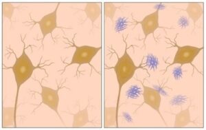
Classification and segmentation of dendritic cells – Results


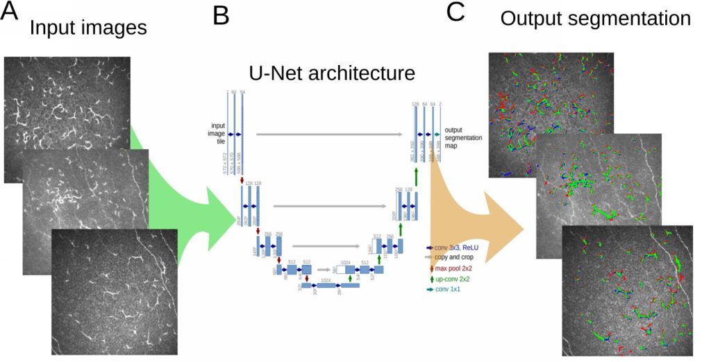
We have obtained 97% accuracy using our U-Net and demonstrated that deep learning can be utilized in the analyses of laser IVCM images, allowing a standardized, objective, and rapid evaluation of ocular surface inflammation. In the attached animated movie, we show an example of IVCM images, an overlay of the U-Net segmentation results (false negative – red, true positive – green, false positive – blue) and ground truth segmentation (white), which are in excellent agreement. Using the automated segmentation, a rapid evaluation of the spatial distribution and density of Dendritic Cells can be given with high accuracy.

Classification and segmentation of dendritic cells

RSIP Vision is a global driving force in the development and implementation of tailor-made solutions in the domain of image processing, computer vision and machine learning. To learn more about RSIP Vision’s activity and view our selected projects, please visit our project pages; to discuss solutions for your project, get directly in touch with our engineering consultants.
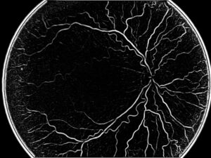
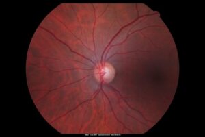
Share

Share on linkedin
Share on twitter
Share on facebook

Main Field

Ophthalmology

RSIP Vision has developed countless projects in the field of ophthalmology for its clients. Computer vision with sophisticated ophthalmic imaging, measurement techniques and AI can yield better results: great precision, accurate diagnosis and best interventional treatment for many pathologies.

View Ophthalmology

Categories

  • Microscopy, Ophthalmology, RSIP Vision Learns

Related Content

Zoom-in-Net

Deep Learning in Ophthalmology

Alzheimer's Disease - AD

Degenerative Diseases Detection in the Eye

Temporary pediatric strabismus in newborn baby

Image processing for pediatric strabismus

ROP - Vessel tortuosity in Retinopathy of Prematurity

ROP: Retinopathy of Prematurity

Eyelid Drooping - MRD1 and MRD2

Eyelid Drooping – Blepharoptosis

Fundus image

Retinal images enhancement

Show all

RSIP Vision

Field-tested software solutions and custom R&D, to power your next medical products with innovative AI and image analysis capabilities.

Read more about us

Get in touch

Please fill the following form and our experts will be happy to reply to you soon

Recent News

PR – Intra-op Virtual Measurements in Laparoscopic and Robotic-Assisted Surgeries

PR – Non-Invasive Planning of Coronary Intervention

PR – Bladder Panorama Generator and Sparse Reconstruction Tool

PR – Registration Module for Orthopedic Surgery

All news
Upcoming Events
Stay informed for our next events
Subscribe to Our Magazines

Subscribe now and receive the Computer Vision News Magazine every month to your mailbox

 
Subscribe for free
Follow us
Linkedin Twitter Facebook Youtube

contact@rsipvision.com

Terms of Use

Privacy Policy

© All rights reserved to RSIP Vision 2021

Created by Shmulik

  • Our Work
    • title-1
      • Ophthalmology
      • Uncategorized
      • Ophthalmology
      • Pulmonology
      • Cardiology
      • Orthopedics
    • Title-2
      • Orthopedics
  • Success Stories
  • Insights
  • The company