Skip to content
  • Our Work
    • Fields
      • Cardiology
      • ENT
      • Gastro
      • Orthopedics
      • Ophthalmology
      • Pulmonology
      • Surgical Intelligence
      • Surgical Robotics
      • Urology
      • Other
    • Modalities
      • Endoscopy
      • Medical Segmentation
      • Microscopy
      • Ultrasound
  • Success Stories
  • Insights
    • Magazine
    • Upcoming Events
    • Webinars
    • Meetups
    • News
    • Blog
  • The company
    • About us
    • Careers
Menu
  • Our Work
    • Fields
      • Cardiology
      • ENT
      • Gastro
      • Orthopedics
      • Ophthalmology
      • Pulmonology
      • Surgical Intelligence
      • Surgical Robotics
      • Urology
      • Other
    • Modalities
      • Endoscopy
      • Medical Segmentation
      • Microscopy
      • Ultrasound
  • Success Stories
  • Insights
    • Magazine
    • Upcoming Events
    • Webinars
    • Meetups
    • News
    • Blog
  • The company
    • About us
    • Careers
Contact

Automatic Pupil Distance Measurement

Automatically Measuring Pupil Distance

For the manufacturing and prescription of modern eye glasses there is a need for high precision measurements. The measured morphological features of the patient’s face and eyes are correlated with physical features of the glasses to ensure the best fit. One of the most important measurements is the interpupillary distance (PD), which is the distance between the projections of the pupil on the cornea (front side of the eye) for the left and right eyes. The center of vision corresponding to the pupil need to be determined with high precision.

Pupil Distance Measurement

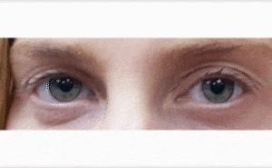
Classical pupil distance measurement methods included the use of a ruler and a pupilmeter. However, these methods were highly error prone and did not fit modern requirements for precision. Such requirements are especially strict in modern glasses, which can accommodate several types of vision corrections: far, intermediate, and near on the same pair. To fit modern precision requirements, lens manufactures have developed systems to measure pupil distance with an error tolerance in the range of one millimeter or less. These measurements are performed on images of the patient’s face, taken while they stand still. However, these systems have several disadvantages, primarily caused by their high cost and lack of portability, due to their large size. As algorithms have advanced some manufacturers have employed expensive 3D technologies for precise pupil distance measurement. Within these systems, patient motion compensation is already embedded, and errors of less than 0.25 millimeters have been achieved. However, these expensive technologies are not yet accessible equally, while vision impairment is frequent throughout the world.While the portability of heavy equipment is limited, and sophisticated technologies are still in the reach of few, a remedy might be the use of portable devices, such as cellphones and tablets. Micro-optics embedded within phones keep improving, and algorithms for pupil distance measurement can be embedded as applications in them.
Sophisticated algorithms developed by RSIP Vision for pupil distance measurement are well-suited for integration into machinery used in eye clinics and can also be embedded as an application in portable devices, such as phones.
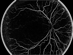
Share

Share on linkedin
Share on twitter
Share on facebook

Main Field

Ophthalmology

RSIP Vision has developed countless projects in the field of ophthalmology for its clients. Computer vision with sophisticated ophthalmic imaging, measurement techniques and AI can yield better results: great precision, accurate diagnosis and best interventional treatment for many pathologies.

View Ophthalmology

Categories

  • Ophthalmology, RSIP Vision Learns

Related Content

Zoom-in-Net

Deep Learning in Ophthalmology

Classification and Segmentation of Dendritic cells

Classification and Segmentation of Dendritic Cells

Alzheimer's Disease - AD

Degenerative Diseases Detection in the Eye

Temporary pediatric strabismus in newborn baby

Image processing for pediatric strabismus

ROP - Vessel tortuosity in Retinopathy of Prematurity

ROP: Retinopathy of Prematurity

Eyelid Drooping - MRD1 and MRD2

Eyelid Drooping – Blepharoptosis

Show all

RSIP Vision

Field-tested software solutions and custom R&D, to power your next medical products with innovative AI and image analysis capabilities.

Read more about us

Get in touch

Please fill the following form and our experts will be happy to reply to you soon

Recent News

PR – Intra-op Virtual Measurements in Laparoscopic and Robotic-Assisted Surgeries

PR – Non-Invasive Planning of Coronary Intervention

PR – Bladder Panorama Generator and Sparse Reconstruction Tool

PR – Registration Module for Orthopedic Surgery

All news
Upcoming Events
Stay informed for our next events
Subscribe to Our Magazines

Subscribe now and receive the Computer Vision News Magazine every month to your mailbox

 
Subscribe for free
Follow us
Linkedin Twitter Facebook Youtube

contact@rsipvision.com

Terms of Use

Privacy Policy

© All rights reserved to RSIP Vision 2021

Created by Shmulik

  • Our Work
    • title-1
      • Ophthalmology
      • Uncategorized
      • Ophthalmology
      • Pulmonology
      • Cardiology
      • Orthopedics
    • Title-2
      • Orthopedics
  • Success Stories
  • Insights
  • The company