Skip to content
  • Our Work
    • Fields
      • Cardiology
      • ENT
      • Gastro
      • Orthopedics
      • Ophthalmology
      • Pulmonology
      • Surgical Intelligence
      • Surgical Robotics
      • Urology
      • Other
    • Modalities
      • Endoscopy
      • Medical Segmentation
      • Microscopy
      • Ultrasound
  • Success Stories
  • Insights
    • Magazine
    • Upcoming Events
    • Webinars
    • Meetups
    • News
    • Blog
  • The company
    • About us
    • Careers
Menu
  • Our Work
    • Fields
      • Cardiology
      • ENT
      • Gastro
      • Orthopedics
      • Ophthalmology
      • Pulmonology
      • Surgical Intelligence
      • Surgical Robotics
      • Urology
      • Other
    • Modalities
      • Endoscopy
      • Medical Segmentation
      • Microscopy
      • Ultrasound
  • Success Stories
  • Insights
    • Magazine
    • Upcoming Events
    • Webinars
    • Meetups
    • News
    • Blog
  • The company
    • About us
    • Careers
Contact

Eyelid Drooping – Blepharoptosis

Blepharoptosis, also known as ptosis, is a drooping of the upper eyelid causing a narrowing of the palpebral fissure (or palpebral aperture), which is the opening between the eye lids.
Eyelid Drooping - MRD1 and MRD2
Eyelid Drooping measurement – MRD1 (orange) and MRD2 (green)

Eyelid drooping may result from any affection of the nerve supply of the upper eyelid musculature, from a disease of the muscles themselves, from mechanical interference in moving the eyelid due to diabetes, from cataract surgery, from the weight of a tumor and from several other conditions. Eyelid drooping could cause functional and cosmetic problems. When the upper lid and eyelashes droop to the extent that they interfere with the line of sight, that is a functional problem and surgical correction via blepharoplasty may be recommended; surgery can help improving visual quality.

It is convenient to analyze an image (or video) in order to evaluate whether the eyelid interferes with the line of sight, impairing visual quality. This may be important to the patient for non-medical reasons too, since blepharoplasty might be covered by the insurance only in case of decline in vision.

Eyelid drooping evaluation method

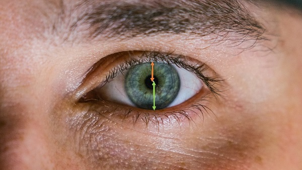
The standard clinical evaluation of ptosis includes manual measurements of vertical distance between the upper and lower lids (with the pupil centered); these are more commonly called MRD1 and MRD2: more specifically, the Margin Reflex Distance 1, defined as the vertical distance between the upper eyelid and the corneal light reflex, and similarly, the Margin Reflex Distance 2, which is the vertical distance between the corneal light reflex and the lower eyelid. The corneal light reflex is the reflection from a light source that is aligned with the visual axis. These measurements quantify the degree of ptosis (which is more severe when palpebral fissure is smaller) and are crucial for the surgical planning in case blepharoplasty is indicated.

Manual measurements are subjective and exposed to human error, as well as time consuming and tedious. Automated image processing is the natural solution for solving these limitations.

Automated eyelid drooping diagnosis by Image Analysis

The automated solution for eyelid drooping is developed using advanced techniques from the computer vision and image processing fields. First, the face must be detected and the corresponding region can be segmented using methods like region growing or edge detection. Region growing involves initial seed selection, therefore the process can be: either semi-automated, by choosing the initial seed points manually; or completely automated, by combining other features from the RGB image that involve similarities between neighbor pixels and dissimilarities with other regions. The eyes are then detected using techniques that use morphological and position features, as well as edge detection algorithm. The corneal light reflex of each eye is identified with the highest-intensity pixel in the histogram of the corresponding eye region or thresholded with a high intensity value. Similarly to what explained above about the manual measurement task, MRD1 and MRD2 are calculated as the vertical distance from the centroid of the corneal light reflex to the edge of the corneal region, which is the eyelid.

These measurement are taken with great precision by the automated eyelid drooping measurement software and the results are available immediately. RSIP Vision has developed a very large portfolio of algorithmic solutions like this, for the benefit of the ophthalmologic community and patients. Read about our projects in image analysis for ophthalmology. You are very welcome to contact our engineers to discuss the best solution for your project.

Contact us now


Share

Share on linkedin
Share on twitter
Share on facebook

Main Field

Ophthalmology

RSIP Vision has developed countless projects in the field of ophthalmology for its clients. Computer vision with sophisticated ophthalmic imaging, measurement techniques and AI can yield better results: great precision, accurate diagnosis and best interventional treatment for many pathologies.

View Ophthalmology

Categories

  • Ophthalmology, RSIP Vision Learns

Related Content

Zoom-in-Net

Deep Learning in Ophthalmology

Classification and Segmentation of Dendritic cells

Classification and Segmentation of Dendritic Cells

Alzheimer's Disease - AD

Degenerative Diseases Detection in the Eye

Temporary pediatric strabismus in newborn baby

Image processing for pediatric strabismus

ROP - Vessel tortuosity in Retinopathy of Prematurity

ROP: Retinopathy of Prematurity

Fundus image

Retinal images enhancement

Show all

RSIP Vision

Field-tested software solutions and custom R&D, to power your next medical products with innovative AI and image analysis capabilities.

Read more about us

Get in touch

Please fill the following form and our experts will be happy to reply to you soon

Recent News

PR – Intra-op Virtual Measurements in Laparoscopic and Robotic-Assisted Surgeries

PR – Non-Invasive Planning of Coronary Intervention

PR – Bladder Panorama Generator and Sparse Reconstruction Tool

PR – Registration Module for Orthopedic Surgery

All news
Upcoming Events
Stay informed for our next events
Subscribe to Our Magazines

Subscribe now and receive the Computer Vision News Magazine every month to your mailbox

 
Subscribe for free
Follow us
Linkedin Twitter Facebook Youtube

contact@rsipvision.com

Terms of Use

Privacy Policy

© All rights reserved to RSIP Vision 2021

Created by Shmulik

  • Our Work
    • title-1
      • Ophthalmology
      • Uncategorized
      • Ophthalmology
      • Pulmonology
      • Cardiology
      • Orthopedics
    • Title-2
      • Orthopedics
  • Success Stories
  • Insights
  • The company