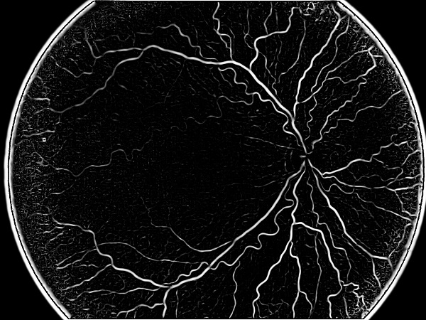
ROP shows no external signs or symptoms. The only way to detect it is through an eye examination by an ophthalmologist. Telemedicine and automated image analysis have the potential to improve access to eye care in less developed and accessible regions, boosting the quality of ROP care.
Computer-based Imaging for Retinopathy of Prematurity diagnosis
Computer-based image analysis has the potential to produce quantifiable, objective measurements.
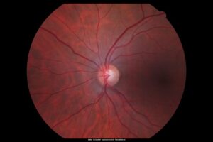
Most approaches for diagnosis include segmentation of retinal vessels and extraction from fundus images of features that account for tortuosity, dilation (diameter) and integrated curvature (sum of angles) for each vessel.
These approaches differ in the various methods used to process the images in order to receive quantifiable values. The first step is pre-processing the images: removing saturation noise by equalizing the histogram of the image and thresholding it to form a binary image. The global threshold value should be adjusted according to the efficiency of thresholding. It is necessary to omit the Optic Disc boundaries before proceeding toward feature extraction. A circular mask can do this job due to the circular morphology of the Optic Disc. Since most ROP features are found close to the optic disc, it is important to define the diagnostic regions at some radius around it to compute features that are relevant to the assessment of the pathology. Next, the vessel centerlines are extracted and a vasculature tree is constructed.
Vessel segmentation approaches are based on algorithmic techniques like matched filter response, multiscale image analysis, morphological segmentation and region growing. For instance, matched filter response measures responses for a binary image with a set of templates in different orientations, based on the assumption that vessels are aligned over the vertical axis and vessels gradients are symmetrical. The response is the outcome of convolution and only the maximum response for each pixel, which indicates of the best match, is retained. Finally, vessel post-processing takes place and measurements are taken for all desired features.
RSIP Vision has a very large portfolio of ophthalmology image analysis projects. This R&D and consulting experience can certainly contribute to the success of your project. Talk about it with our engineers!

 Ophthalmology