Skip to content
  • Our Work
    • Fields
      • Cardiology
      • ENT
      • Gastro
      • Orthopedics
      • Ophthalmology
      • Pulmonology
      • Surgical Intelligence
      • Surgical Robotics
      • Urology
      • Other
    • Modalities
      • Endoscopy
      • Medical Segmentation
      • Microscopy
      • Ultrasound
  • Success Stories
  • Insights
    • Magazine
    • Upcoming Events
    • Webinars
    • Meetups
    • News
    • Blog
  • The company
    • About us
    • Careers
Menu
  • Our Work
    • Fields
      • Cardiology
      • ENT
      • Gastro
      • Orthopedics
      • Ophthalmology
      • Pulmonology
      • Surgical Intelligence
      • Surgical Robotics
      • Urology
      • Other
    • Modalities
      • Endoscopy
      • Medical Segmentation
      • Microscopy
      • Ultrasound
  • Success Stories
  • Insights
    • Magazine
    • Upcoming Events
    • Webinars
    • Meetups
    • News
    • Blog
  • The company
    • About us
    • Careers
Contact

Computer-Assisted Joint Replacement (hip and knee)

Pathologies in the joint regions are common especially among elderly patients. They are caused, in many cases, by wear and tear of the cartilage layer cushioning between bones. The consequences of this deterioration can be serious and affect the proper function of the joint. Cases where the hip and knee begin to lose cartilage are very frequent and often require treatment.


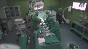
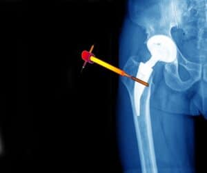
One effective way of dealing with joint deterioration is by undergoing surgical replacement. Hip replacement (Total Hip Arthroplasty or THA) and knee replacement (Total Knee Arthroplasty or TKA) are frequent surgical interventions, which are rigorously planned in advance, a process which involves accurate measurements of various anatomical locations. A Computer Tomography (CT) image of the operative area is taken and a patient-specific model of the bone is prepared. The artificial joint is built upon this model, taking into account many patient-specific parameters such as the bone size, structure etc. Accurate planning is key to ensure that the new joint fits the patient anatomy and remains stable over a long period of time.

Computer-Assisted Joint Replacement Software

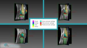
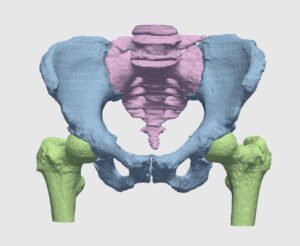
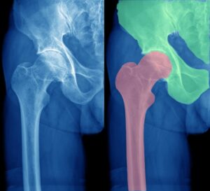
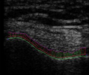
RSIP Vision has developed a software package based on sophisticated computer vision algorithms and the most advanced image processing techniques to aid in the modelling process with a very high anatomic precision. Commonly, the models are as precise as the resolution of the CT allows. Our software performs segmentation of the bones using the CT volume. The main challenge of this segmentation is the difficulty to identify the border between bone and cartilage. We solve this challenge by utilizing the Hounsfield units, gradients and shape structure into an optimization algorithm to trace the edges between soft tissue, cartilage and bones. Our software is able, in just a few minutes, to delimit edges and complete the segmentation, which otherwise would have taken hours for an expert physician to complete. Additionally, anatomical landmarks are detected and utilized in the fitting process. Current work on the next generation of RSIP Vision’s computer-assisted joint replacement software package is based on deep learning techniques, and it shows great results.

Consult our experts at RSIP Vision

Share

Share on linkedin
Share on twitter
Share on facebook

Main Field

Orthopedics

RSIP Vision brings specific field-tested experience in solving real world orthopedic imaging challenges for multiple clinical use cases. From preoperative planning, to intraoperative navigation, and many more. We are a proven leader, enabling our clients to bring innovative AI capabilities into the market.

View Orthopedics

Categories

  • Orthopedics, RSIP Vision Learns

Related Content

Intraoperative Registration Module for Orthopedic Surgery

PR – Registration Module for Orthopedic Surgery

Automated Assessment of Cartilage Damage

PR – Automated Assessment of Cartilage Damage

Hip

Segmentation in Orthopedics with Deep Learning

Surgery tool as seen on the CT scan

Navigation Systems in Orthopedic Surgery

Orthopedic Surgery - CT Segmentation

CT Segmentation in orthopedic surgery

Ultrasound Imaging of Speckled Cartilage Tissue

Density Measurement of Damaged Cartilage

Show all

RSIP Vision

Field-tested software solutions and custom R&D, to power your next medical products with innovative AI and image analysis capabilities.

Read more about us

Get in touch

Please fill the following form and our experts will be happy to reply to you soon

Recent News

PR – Intra-op Virtual Measurements in Laparoscopic and Robotic-Assisted Surgeries

PR – Non-Invasive Planning of Coronary Intervention

PR – Bladder Panorama Generator and Sparse Reconstruction Tool

PR – Registration Module for Orthopedic Surgery

All news
Upcoming Events
Stay informed for our next events
Subscribe to Our Magazines

Subscribe now and receive the Computer Vision News Magazine every month to your mailbox

 
Subscribe for free
Follow us
Linkedin Twitter Facebook Youtube

contact@rsipvision.com

Terms of Use

Privacy Policy

© All rights reserved to RSIP Vision 2021

Created by Shmulik

  • Our Work
    • title-1
      • Ophthalmology
      • Uncategorized
      • Ophthalmology
      • Pulmonology
      • Cardiology
      • Orthopedics
    • Title-2
      • Orthopedics
  • Success Stories
  • Insights
  • The company