Skip to content
  • Our Work
    • Fields
      • Cardiology
      • ENT
      • Gastro
      • Orthopedics
      • Ophthalmology
      • Pulmonology
      • Surgical Intelligence
      • Surgical Robotics
      • Urology
      • Other
    • Modalities
      • Endoscopy
      • Medical Segmentation
      • Microscopy
      • Ultrasound
  • Success Stories
  • Insights
    • Magazine
    • Upcoming Events
    • Webinars
    • Meetups
    • News
    • Blog
  • The company
    • About us
    • Careers
Menu
  • Our Work
    • Fields
      • Cardiology
      • ENT
      • Gastro
      • Orthopedics
      • Ophthalmology
      • Pulmonology
      • Surgical Intelligence
      • Surgical Robotics
      • Urology
      • Other
    • Modalities
      • Endoscopy
      • Medical Segmentation
      • Microscopy
      • Ultrasound
  • Success Stories
  • Insights
    • Magazine
    • Upcoming Events
    • Webinars
    • Meetups
    • News
    • Blog
  • The company
    • About us
    • Careers
Contact

Navigation Systems in Orthopedic Surgery

The navigation system is one of the most powerful tools for the surgeon in orthopedic surgery. The visualization system aids the surgeon to place his or her tools without excessive exposure to X-ray radiations.

Orthopedic Surgery Navigation System

The objectives of the Navigation System

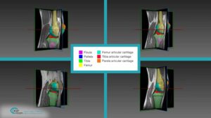
  • Present 2D and 3D views of the surgery location in the 3D CT scan
  • Adjust the view orientation to match the tool’s orientation (or derived standard orientations)
  • Real time performance – minimize display delays to allow high accuracy navigation


Real-Time Navigation System – Procedure

The navigation system uses a real-time transformation sent by the tracking system. This transformation contains the surgeon’s tool’s tip and its orientation. The transformation is being updated at a high rate (about 25-30 frame per second) – following very closely to the tool’s movements.

Multiple registrations, performed in the preceding phases, supply the mapping from the tracking system’s “world” to the 3D CT “world”. Using this transformation, the algorithm calculates the location and the orientation of the tool’s tip in the 3D CT scans. The display system extracts and displays the views (2D and 3D), as previously defined by the surgeon.

The tracking system monitors the tool’s location at a high rate. The transformation of the tool’s location and orientation is calculated at the same pace, letting the display adjust smoothly.

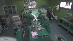
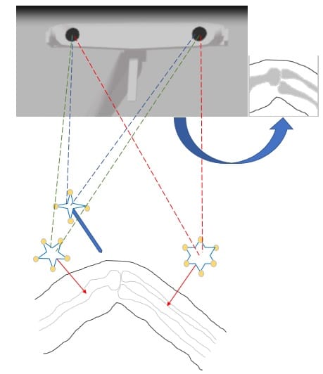
A typical tracking system is based on hardware (markers attached to key objects and cameras) and software (constructing a 3D model of the scene).

Markers can be passive or active, where an active marker is one that releases a signal to indicate its location and the passive marker is constructed of well-defined objects (spheres) that are easily detected. Cameras can be RGB or IR, like passive system cameras or any sensor able to receive signals coming from active sensors.

The system uses at least two cameras (or sensors) and it reconstructs the 3D scene employing stereo vision methods. The cameras are positioned at some distance in between, so that each one can “see” the markers from a slightly different angle of view through a depth map with accurate location of each marker in the 3D space. Each marker is constructed of a unique shape or pattern of objects. It enables the detection and classification algorithm to identify each of the marker’s identifications. This feature is important because each marker is attached to a different object.

Markers in Orthopedic navigation system

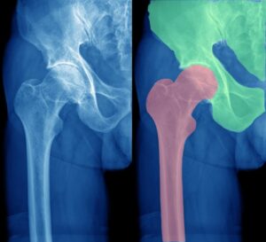
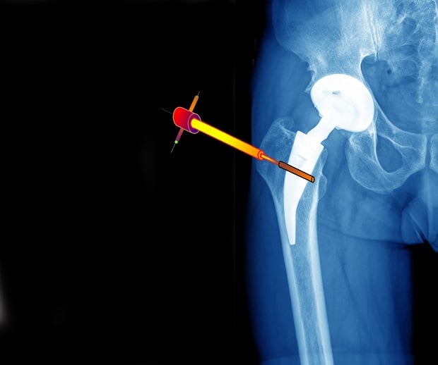
The tracking system is part of the in-operation navigation system. Markers are attached to key objects like the bones or the surgeon’s tools. The navigation task logs the patient orientation and positioning as well as the tool’s momentarily location, while the navigation system exploits this information to “navigate” inside the 3D patient’s CT according to the tools’ location and orientation. The pre-operation phase generates the required transformation between the CT scan and the tracking coordinate system (real world coordinates). Using this transformation matrix, each new tool’s location and orientation is translated into a new view of the CT, visible to the surgeon on the specific display system.

This is the forth in a series of five articles about Orthopedic Navigation. Read the next article about Validation of Navigation System’s Accuracy in Orthopedic Surgeries. You can also read about RSIP Vision’s projects in Orthopedic Surgery.

Share

Share on linkedin
Share on twitter
Share on facebook

Main Field

Orthopedics

RSIP Vision brings specific field-tested experience in solving real world orthopedic imaging challenges for multiple clinical use cases. From preoperative planning, to intraoperative navigation, and many more. We are a proven leader, enabling our clients to bring innovative AI capabilities into the market.

View Orthopedics

Categories

  • Orthopedics, RSIP Vision Learns

Related Content

Intraoperative Registration Module for Orthopedic Surgery

PR – Registration Module for Orthopedic Surgery

Automated Assessment of Cartilage Damage

PR – Automated Assessment of Cartilage Damage

Hip

Segmentation in Orthopedics with Deep Learning

Knee

Computer-Assisted Joint Replacement (hip and knee)

Orthopedic Surgery - CT Segmentation

CT Segmentation in orthopedic surgery

Ultrasound Imaging of Speckled Cartilage Tissue

Density Measurement of Damaged Cartilage

Show all

RSIP Vision

Field-tested software solutions and custom R&D, to power your next medical products with innovative AI and image analysis capabilities.

Read more about us

Get in touch

Please fill the following form and our experts will be happy to reply to you soon

Recent News

PR – Intra-op Virtual Measurements in Laparoscopic and Robotic-Assisted Surgeries

PR – Non-Invasive Planning of Coronary Intervention

PR – Bladder Panorama Generator and Sparse Reconstruction Tool

PR – Registration Module for Orthopedic Surgery

All news
Upcoming Events
Stay informed for our next events
Subscribe to Our Magazines

Subscribe now and receive the Computer Vision News Magazine every month to your mailbox

 
Subscribe for free
Follow us
Linkedin Twitter Facebook Youtube

contact@rsipvision.com

Terms of Use

Privacy Policy

© All rights reserved to RSIP Vision 2021

Created by Shmulik

  • Our Work
    • title-1
      • Ophthalmology
      • Uncategorized
      • Ophthalmology
      • Pulmonology
      • Cardiology
      • Orthopedics
    • Title-2
      • Orthopedics
  • Success Stories
  • Insights
  • The company