Skip to content
  • Our Work
    • Fields
      • Cardiology
      • ENT
      • Gastro
      • Orthopedics
      • Ophthalmology
      • Pulmonology
      • Surgical Intelligence
      • Surgical Robotics
      • Urology
      • Other
    • Modalities
      • Endoscopy
      • Medical Segmentation
      • Microscopy
      • Ultrasound
  • Success Stories
  • Insights
    • Magazine
    • Upcoming Events
    • Webinars
    • Meetups
    • News
    • Blog
  • The company
    • About us
    • Careers
Menu
  • Our Work
    • Fields
      • Cardiology
      • ENT
      • Gastro
      • Orthopedics
      • Ophthalmology
      • Pulmonology
      • Surgical Intelligence
      • Surgical Robotics
      • Urology
      • Other
    • Modalities
      • Endoscopy
      • Medical Segmentation
      • Microscopy
      • Ultrasound
  • Success Stories
  • Insights
    • Magazine
    • Upcoming Events
    • Webinars
    • Meetups
    • News
    • Blog
  • The company
    • About us
    • Careers
Contact

Density Measurement of Damaged Cartilage

Knee cartilage acts as a shock absorber, useful to absorb and damp shock impulses between the thigh (femur) and the shin (tibia) bones. The cartilage layer is a flexible tissue, which serves to ease the pressure put on the knee joint caused by mere walking and the weight of the body it has to carry. Naturally, the cartilage is eroded over the years (osteoarthritis). Moreover, the cartilage layer can be damaged or injured also by accidents. For severe knee cartilage damage, a clinical intervention is needed, such as drilling small holes (to encourage growth of new cartilage), cartilage implant, gel injection, and in severe cases a total replacement of the knee cartilage.
Ultrasound Imaging of Speckled Cartilage Tissue
Ultrasound imaging of speckled cartilage tissue

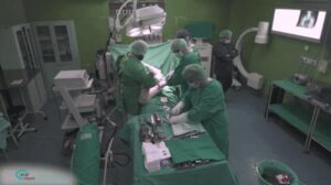
The extent of the intervention is determined by physicians based on the current erosion state. The state of erosion can be observed by the way of non-invasive imaging techniques such as simple ultrasonic imaging. Trained orthopedics evaluate the level of the intervention needed and steer the course of action based on, among other things, the thickness of the knee cartilage, as assessed by the physician. In fact, output images are analyzed and measured by hand or with semi-automated computational tools.

Automated density measurement of damaged cartilage

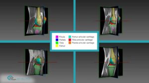
RSIP Vision has developed an automated tool to measure knee cartilage thickness based on ultrasonic images. Our automated algorithmic procedure releases physicians from the time-consuming human assessment. Ultrasound images of cartilage are hard to work with since borders between tissues are fuzzy. These boundaries take a cloud-like form, which can morph in both density and texture. To ensure automatic and accurate recognition of the different textures, we utilized active contour – ‘snakes’ technology to determine the borders of the cartilage. Our density measurement of damaged cartilage software is used by medical staff to produce a non-invasive, efficient and accurate measurement of knee cartilage density from ultrasound imaging of speckled cartilage tissue.

When shock absorption properties of cartilage are reduced by damage, RSIP Vision’s software enables orthopedics to assess state of erosion and decide treatment. To learn more about the activity of RSIP Vision in non-invasive orthopedic surgery, please visit our orthopedic surgery and medical segmentation project pages.

Share

Share on linkedin
Share on twitter
Share on facebook

Main Field

Orthopedics

RSIP Vision brings specific field-tested experience in solving real world orthopedic imaging challenges for multiple clinical use cases. From preoperative planning, to intraoperative navigation, and many more. We are a proven leader, enabling our clients to bring innovative AI capabilities into the market.

View Orthopedics

Categories

  • Orthopedics, RSIP Vision Learns

Related Content

Intraoperative Registration Module for Orthopedic Surgery

PR – Registration Module for Orthopedic Surgery

Automated Assessment of Cartilage Damage

PR – Automated Assessment of Cartilage Damage

Hip

Segmentation in Orthopedics with Deep Learning

Knee

Computer-Assisted Joint Replacement (hip and knee)

Surgery tool as seen on the CT scan

Navigation Systems in Orthopedic Surgery

Orthopedic Surgery - CT Segmentation

CT Segmentation in orthopedic surgery

Show all

RSIP Vision

Field-tested software solutions and custom R&D, to power your next medical products with innovative AI and image analysis capabilities.

Read more about us

Get in touch

Please fill the following form and our experts will be happy to reply to you soon

Recent News

PR – Intra-op Virtual Measurements in Laparoscopic and Robotic-Assisted Surgeries

PR – Non-Invasive Planning of Coronary Intervention

PR – Bladder Panorama Generator and Sparse Reconstruction Tool

PR – Registration Module for Orthopedic Surgery

All news
Upcoming Events
Stay informed for our next events
Subscribe to Our Magazines

Subscribe now and receive the Computer Vision News Magazine every month to your mailbox

 
Subscribe for free
Follow us
Linkedin Twitter Facebook Youtube

contact@rsipvision.com

Terms of Use

Privacy Policy

© All rights reserved to RSIP Vision 2021

Created by Shmulik

  • Our Work
    • title-1
      • Ophthalmology
      • Uncategorized
      • Ophthalmology
      • Pulmonology
      • Cardiology
      • Orthopedics
    • Title-2
      • Orthopedics
  • Success Stories
  • Insights
  • The company