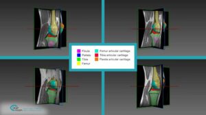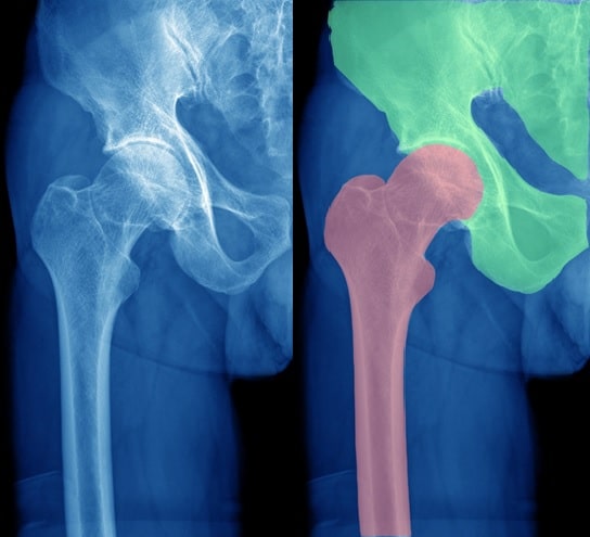
The objectives of CT segmentation in orthopedic surgery are:
- Detect bones in 3D CT or 2D C-Arm imaging
- Convert the detected object from voxel (pixel) related to its outer (more compact) mesh representation.
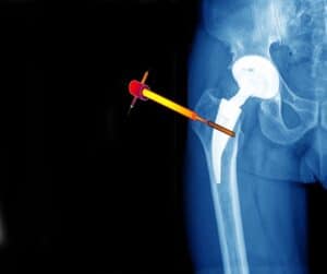
- Make precise measurements for the placement of the implant
Procedure
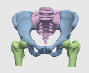
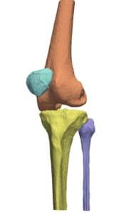
The CT scan are first localized. Each of the bones contained in it is bounded. The bounding box or surface is used to specify the location (in 3D or 2D) where the bone is found. The segmentation algorithm works on each bone separately. It detects the contained voxels (pixels). Several algorithmic techniques are used, starting from region growing, shape classifiers, parametric modeling or any combination of them. What follows will help us understand the state-of-the-art techniques for CT segmentation in orthopedic surgery.
Cheng Li – “A new algorithm for cortical bone segmentation with its validation and applications to in vivo imaging” presents a new cortical bone segmentation method that utilizes larger contextual and topologic knowledge of distal tibia bone through fuzzy distance transform and connectivity analysis
J. Zhang, C.-H. Yan, C.-K. Chui, S.-H. Ong – “Fast segmentation of bone in CT images using 3D adaptive thresholding” propose an automatic, fast, robust and accurate method for the segmentation of bone using 3D adaptive thresholding. An initial segmentation is first performed to partition the image into bone and non-bone classes, followed by an iterative process of 3D correlation to update voxel classification. This iterative process significantly improves the thresholding performance.
The Magic Wand algorithm is a semi-automatic method for image segmentation. The user locates a point which lays inside the area to be segmented. The algorithm calculates statistics for this location. A region growing is performed on surrounding pixels which match the statistics up to some (user-controlled) tolerance. This solution is used to segment bones from tissues due to their different gray levels in CT scan. However, in cases where the bones density is not uniform, a few iterations may be required until acceptable segmentation is achieved. The Magic Wand method gives a good initial solution for bones segmentation.
Graph-Cuts algorithm was once considered a satisfying technology for medical segmentation. The method is based on Graph Theory applied to image segmentation. Each pixel of the image is a node in the graph. CT images of bones and tissues are labeled as objects and background. The Max-Flow/Min-Cut algorithm is used for optimal pixel labelling. The algorithm is started by getting location(s) of bones and background from the user. The optimal labeling is based on high gradients – location of the bones contours. The downfall of this algorithm is that it does not consider information like bones shape. In addition, it requires a good seed. For this and many other reasons, Graph cuts was never the only chosen method; today, AI techniques like deep learning are widely used and recommended for CT segmentation in orthopedic surgery.
Deep learning is constantly being proven to be the most powerful tool in various tasks, with segmentation as no exception. The medical field has gained significant progress by utilizing deep learning techniques. Generic, out-of-the-box solutions exist, and can produce fair results. However, in order to make the most out of a deep learning solution, multiple consideration should be taken into account. For instance, proper architecture, correct input and proper constraints can significantly boost the results. We choose this solution for almost every segmentation project. Metal artifacts simulation and specific bone emphasis were very beneficial for the bone segmentation task. The platform used for this work was our own developed variant of U-Net with shape-aware loss. Thus, our solution makes wide use of Convolutional Neural Netwoks (CNN). Even though unsupervised techniques are available, best performing algorithms require annotated data. To train this world-class AI, we first annotated multiple scans via our proprietary algorithm for initial bone segmentation; this was based on bone geometry, gradients and texture. After that phase, our trained medical image analysts examined and further improved the annotations, working hand in hand with expert radiologists.
This is the second in a series of five articles about Orthopedic Navigation. Read the next article about Registration of CT and X-Ray in Orthopedic Surgeries.
 Orthopedics