Skip to content
  • Our Work
    • Fields
      • Cardiology
      • ENT
      • Gastro
      • Orthopedics
      • Ophthalmology
      • Pulmonology
      • Surgical Intelligence
      • Surgical Robotics
      • Urology
      • Other
    • Modalities
      • Endoscopy
      • Medical Segmentation
      • Microscopy
      • Ultrasound
  • Success Stories
  • Insights
    • Magazine
    • Upcoming Events
    • Webinars
    • Meetups
    • News
    • Blog
  • The company
    • About us
    • Careers
Menu
  • Our Work
    • Fields
      • Cardiology
      • ENT
      • Gastro
      • Orthopedics
      • Ophthalmology
      • Pulmonology
      • Surgical Intelligence
      • Surgical Robotics
      • Urology
      • Other
    • Modalities
      • Endoscopy
      • Medical Segmentation
      • Microscopy
      • Ultrasound
  • Success Stories
  • Insights
    • Magazine
    • Upcoming Events
    • Webinars
    • Meetups
    • News
    • Blog
  • The company
    • About us
    • Careers
Contact

Coronary CT Angiography with Deep Learning

Coronary artery pathologies are a leading cause of heart failure and death in the western world. Narrowing and hardening of the vessels carrying oxygen-rich blood to the heart, due to the accumulation of plaque on the vascular walls, is a major source of atherosclerosis, a major cardiovascular disease. The accumulation of plaque on the capillary and artery walls disrupts permeability and pressure leading to reduced ability of the heart to pump blood to distal body parts. Such abnormalities are manifested in symptoms such as arrhythmia, angina, appearance of blood clots, and finally heart attack.

Coronary CT Angiography

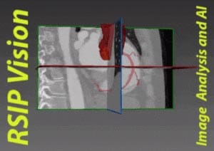
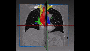
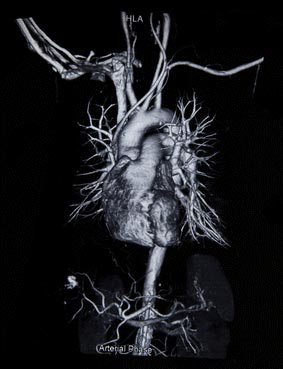
The health of coronary tree is monitored in clinics using 3D computed tomography (CT) angiography and ultrasound. Using CT, a 3-dimensional model of the heart and its vicinity can be constructed by collecting the slices obtained. Angiography enables visualization of the blood flow in arteries and capillaries, hence it is used as a method to visualize the blood vessels. Narrowing of the blood vessels can be located by CT angiography, thus allowing planning of invasive clinical intervention, such as stent placement.

Clinical intervention in pathological cases requires careful planning. A computerized model of the blood flow serves as a navigation, investigation and analysis tool before the intervention. However, manual inspection of the resulting image hinders timely response. The complexity of the vascular tree seen in the 3D representation slows down significantly the detection of narrowed regions, leaky vessels and other pathologies.

Automated detection of anomalies on coronary CT angiography using deep learning

A computational approach to the detection of anomalies can therefore prove to be superior to manual labor, both in terms of processing time and accurate localization of pathologies. Since the deep learning revolution, the algorithmic approach produces state-of-the-art results, much better than with any previous method.

For this end, RSIP Vision has constructed an algorithm to detect coronary anomalies from CT image stacks, which automatically segments the arteries. The production of 3D CT images of the active heart requires fast imaging technology which works on multiple scanned layers simultaneously. The main challenge for our software was to automatically separate the different components of the 3D image. We needed to locate the muscular layer (myocardium) of the heart in the images to guide the rest of the segmentation process.

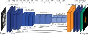
Prior to the training, the data set was cleaned, and problematic scans were removed . We generated the training dataset for our Convolutional Neural Network (CNN) using traditional computer vision techniques: namely, ‘minimal graph cut’, an algorithmic image segmentation technique taken from the graph theory world, to separate the myocardium from the visceral and parietal pericardium layers of the heart within the images. Our U-Net based CNN was trained with these labeled data. The results of this cutting edge deep learning procedure are far better than with any of the methods that were available beforehand.

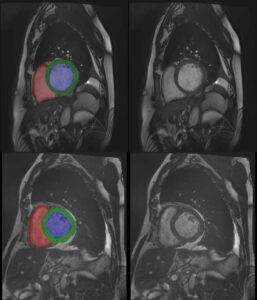
The software is used to accurately segment arteries, locate blockages and pinpoint other abnormalities. This non-invasive process suppresses the need for (sometime dangerous) cardiac catheterization procedures, sparing thus needless risks of injuries. RSIP Vision has been specializing in cardiological surgery computational aids for the past 20 years. Our deep learning algorithms, put to challenge in both sterile and clinical applications, have proven extremely successful in coronary CT angiography anomalies detection and in all kinds of image processing in cardiology tasks. Take us along for your next Deep Learning project.

Share

Share on linkedin
Share on twitter
Share on facebook

Main Field

Cardiology

RSIP Vision’s advanced algorithms and image processing software (together with proper machines and devices) are used by physicians and researchers to develop and perform preventive and interventional cardiovascular procedures: that's our contribution to Artificial Intelligence in cardiology. Our software provides information about the patient’s cardiovascular system in a non-invasive manner, helps physicians ensure accuracy during complex operations, raises the level of patient safety through limiting his/her exposure to radiation, and aids diagnosis. We come to the office to write algorithms that save lives. In this section about computer vision in cardiology you can discover some of the ways we do it.

View Cardiology

Categories

  • Cardiology, RSIP Vision Learns

Related Content

PR – Non-Invasive Planning of Coronary Intervention

AI in Cardiac CT angiography

Coronary Arteries Segmentation

Great Vessels Segmentation with Deep Learning

Cardiac Motion Correction

Deep Learning in Cardiology

Cardiac MRI 3D automatic segmentation

3D Cardiac MRI automatic segmentation

PR – Non-Invasive Planning of Coronary Intervention

AI in Cardiac CT angiography

Coronary Arteries Segmentation

Great Vessels Segmentation with Deep Learning

Cardiac Motion Correction

Deep Learning in Cardiology

Cardiac MRI 3D automatic segmentation

3D Cardiac MRI automatic segmentation

Show all

RSIP Vision

Field-tested software solutions and custom R&D, to power your next medical products with innovative AI and image analysis capabilities.

Read more about us

Get in touch

Please fill the following form and our experts will be happy to reply to you soon

Recent News

PR – Intra-op Virtual Measurements in Laparoscopic and Robotic-Assisted Surgeries

PR – Non-Invasive Planning of Coronary Intervention

PR – Bladder Panorama Generator and Sparse Reconstruction Tool

PR – Registration Module for Orthopedic Surgery

All news
Upcoming Events
Stay informed for our next events
Subscribe to Our Magazines

Subscribe now and receive the Computer Vision News Magazine every month to your mailbox

 
Subscribe for free
Follow us
Linkedin Twitter Facebook Youtube

contact@rsipvision.com

Terms of Use

Privacy Policy

© All rights reserved to RSIP Vision 2021

Created by Shmulik

  • Our Work
    • title-1
      • Ophthalmology
      • Uncategorized
      • Ophthalmology
      • Pulmonology
      • Cardiology
      • Orthopedics
    • Title-2
      • Orthopedics
  • Success Stories
  • Insights
  • The company