Fundus images enhancement
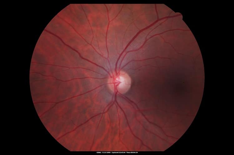
It is highly recommended to use image histogram stretching techniques in fundus images. Information about the distribution of grey levels in a specific image is provided by the image histogram. In an exceptionally dark or bright image, the grey level would be clustered to the extremes of the histogram, but in a well-contrasted image these levels would be well spread over a wide range. In low contrast image, the solution is given by histogram stretching algorithms: these algorithms are able to distribute grey levels more equally across the range, according to specific user-defined equations, and therefore produce an image with greater contrast than the original. Histogram equalization is a basic automated process, which aims at making the histogram uniformly distributed. When using an adaptive Histogram Equalization method, the image is divided to sub-images, each having a sub-histogram which is then stretched. The neighboring sub-images are stitched together using bilinear interpolation to eliminate the boundaries between them.
Other retinal images enhancement methods such as filtering make the image more suitable for use: one of these filtering methods is averaging, that is able to reduce noise; another filtering method involves boundaries enhancement.
OCT enhancement
OCT is extremely useful in assessing optic nerve damages in glaucoma, retinal detachment and macular edema. Evaluation of these pathologies requires assessment of changes over time, which might be very hard to tell in some images. OCT images are subject to various distortions that introduce artifacts in the OCT cross-sections such as floaters, saccades and speckle noise.
OCT retinal image enhancement methods are different from those which we have seen giving such good results on fundus images: signal averaging is one of the most important and effective techniques to improve OCT image quality. This technique has practical limitations due to the inability of patients to maintain fixation during examination and it requires hardware eye-tracking, which is not necessarily available in every clinic using OCT. Due to eye movement (eye saccades, blinks and patient’s movement) there is a limitation on the number of frames that can be recorded and averaged.
For that reason, we recommend using an enhancement method by virtually averaging OCT signal (no need for eye tracking). The technique uses Gaussian random distribution for each sampling voxel on a 3x3x3 cube of neighboring voxels; this is repeated an appropriate amount of times such as ensures the optimization of the averaging result.
Both signal to noise ratio (SNR) and contrast to noise ratio (CNR) are improved by this method: these parameters indicate that the non-averaged non-tracking OCT image quality is significantly better.
Another denoising technique consists in a locally adaptive denoising algorithm using a combination of the Double-Density Discrete Wavelet Transform (DDDWT) and the Dual-Tree Complex Wavelet Transform (DTCWT), useful to reduce speckle noise in OCT images of the retina. The algorithm overcomes the limitations of commonly used multiple frame averaging technique, improving SNR.
RSIP Vision has been at the forefront of these retinal images enhancement techniques for several decades. Coupled with an excellent knowledge of the eye’s physiology, our expertise will have an essential role in the success of your ophthalmology software. Contact our consultants to find out what we can do for you!

 Ophthalmology