Skip to content
  • Our Work
    • Fields
      • Cardiology
      • ENT
      • Gastro
      • Orthopedics
      • Ophthalmology
      • Pulmonology
      • Surgical Intelligence
      • Surgical Robotics
      • Urology
      • Other
    • Modalities
      • Endoscopy
      • Medical Segmentation
      • Microscopy
      • Ultrasound
  • Success Stories
  • Insights
    • Magazine
    • Upcoming Events
    • Webinars
    • Meetups
    • News
    • Blog
  • The company
    • About us
    • Careers
Menu
  • Our Work
    • Fields
      • Cardiology
      • ENT
      • Gastro
      • Orthopedics
      • Ophthalmology
      • Pulmonology
      • Surgical Intelligence
      • Surgical Robotics
      • Urology
      • Other
    • Modalities
      • Endoscopy
      • Medical Segmentation
      • Microscopy
      • Ultrasound
  • Success Stories
  • Insights
    • Magazine
    • Upcoming Events
    • Webinars
    • Meetups
    • News
    • Blog
  • The company
    • About us
    • Careers
Contact

Geographic Atrophy (GA)

Geographic Atrophy (generally called GA) is a case of advanced Dry AMD (Age-related Macular Degeneration) which might lead to vision loss. As a consequence of the thinning of the retina and loss of RPE layer (Retinal Pigment Epithelium, located just outside the retina) via death of rods and cones, islands of lost photoreceptor cells are formed. These are visible in eye care imaging, which allows to detect and quantify the disease.
Geographic Atrophy with Drusen
Geographic Atrophy with calcified macular drusen
At the early stage of this pathology protein deposits called drusen (which were generated at the back of the retina) can appear, disturbing the vision. During the development of the disease, the scattered locations of the drusen gather together leading to the next stage, which is Geographic Atrophy. One of the goals at this point is to monitor and measure the GA, in order to decide the appropriate treatment for each case, according to its effectiveness to stop or at least control the development of the disease.
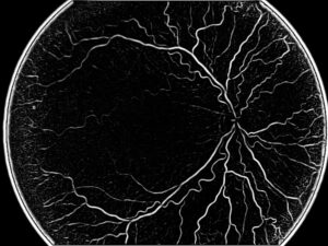
Different imaging methods are used: among them, IR imaging, FAF (fundus autofluorescence), regular fundus images and OCT. Having images coming from all these modalities is the ideal solution, however each one of them can provide good enough pictures to assess the situation.

Geographic Atrophy Segmentation

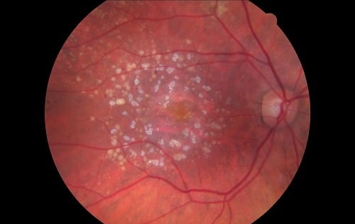
The problem in estimating the size of the GA is that in many cases the boundaries are not very clear. When boundaries are clear, we can use the segmentation methods which are well known in the literature, specifically adjusted as fit for retina analysis. Nevertheless, difficulties are to be expected when the contours of the GA are not so clear-cut, at the three levels: the background of the eye (which can be quite dark); the GA itself, represented by a clear area; and the parts in between, who can also be quite dark, with no certainty whether they belong to the GA or not.
Software has to deal with all these challenges. When the boundaries are clear, all is good. But when boundaries are not clear enough, the results are not sufficient for giving sufficient information to the ophthalmologist whether the area belongs or not to GA. Human evaluation in this task is subject to high inter-observer and intra-observer variation, associating different areas to the Geographic Atrophy at each observation, which makes less reliable the observation of changes over time.
Segmentation in the medical field relies often on the following algorithmic techniques: first and foremost region growing, which in many cases is nearly intuitive and quite easy to find: the darkest point will be associated to GA, within which a seed will be identified and grown until they reach the pixels belonging to the retina itself. Another technique is segmentation based on Otsu, a method designed to find the best separation between two populations, based on the histogram; in this approach, we take the threshold that leaves minimal variance to the two separate groups. One more technique is graph-cuts, which enable to balance segmentation between proximity of the pixels and their gray level. This algorithm is based on the graph theory, finding the minimal cut in a flow graph. Used in image processing, this technology cuts between two levels: source (in this case, the area within the GA) and sink (the retina itself). Its mission is to find the best cut separating the pixels of the source and background.
The seed to be used in region growing can be selected either by human expert or by the machine: when the image is very clear and the Geographic Atrophy has clear boundaries, we can let the algorithms detect the seed without any input by the ophthalmologist. On more ambiguous images, it’s recommended to let the physician control this process.
RSIP Vision has a rich experience in developing algorithmic solutions for many technology companies in the field of ophthalmology. Take advantage of our experience and consult our engineers before starting your project.

Share

Share on linkedin
Share on twitter
Share on facebook

Main Field

Ophthalmology

RSIP Vision has developed countless projects in the field of ophthalmology for its clients. Computer vision with sophisticated ophthalmic imaging, measurement techniques and AI can yield better results: great precision, accurate diagnosis and best interventional treatment for many pathologies.

View Ophthalmology

Categories

  • Ophthalmology, RSIP Vision Learns

Related Content

Zoom-in-Net

Deep Learning in Ophthalmology

Classification and Segmentation of Dendritic cells

Classification and Segmentation of Dendritic Cells

Alzheimer's Disease - AD

Degenerative Diseases Detection in the Eye

Temporary pediatric strabismus in newborn baby

Image processing for pediatric strabismus

ROP - Vessel tortuosity in Retinopathy of Prematurity

ROP: Retinopathy of Prematurity

Eyelid Drooping - MRD1 and MRD2

Eyelid Drooping – Blepharoptosis

Show all

RSIP Vision

Field-tested software solutions and custom R&D, to power your next medical products with innovative AI and image analysis capabilities.

Read more about us

Get in touch

Please fill the following form and our experts will be happy to reply to you soon

Recent News

PR – Intra-op Virtual Measurements in Laparoscopic and Robotic-Assisted Surgeries

PR – Non-Invasive Planning of Coronary Intervention

PR – Bladder Panorama Generator and Sparse Reconstruction Tool

PR – Registration Module for Orthopedic Surgery

All news
Upcoming Events
Stay informed for our next events
Subscribe to Our Magazines

Subscribe now and receive the Computer Vision News Magazine every month to your mailbox

 
Subscribe for free
Follow us
Linkedin Twitter Facebook Youtube

contact@rsipvision.com

Terms of Use

Privacy Policy

© All rights reserved to RSIP Vision 2021

Created by Shmulik

  • Our Work
    • title-1
      • Ophthalmology
      • Uncategorized
      • Ophthalmology
      • Pulmonology
      • Cardiology
      • Orthopedics
    • Title-2
      • Orthopedics
  • Success Stories
  • Insights
  • The company