Skip to content
  • Our Work
    • Fields
      • Cardiology
      • ENT
      • Gastro
      • Orthopedics
      • Ophthalmology
      • Pulmonology
      • Surgical Intelligence
      • Surgical Robotics
      • Urology
      • Other
    • Modalities
      • Endoscopy
      • Medical Segmentation
      • Microscopy
      • Ultrasound
  • Success Stories
  • Insights
    • Magazine
    • Upcoming Events
    • Webinars
    • Meetups
    • News
    • Blog
  • The company
    • About us
    • Careers
Menu
  • Our Work
    • Fields
      • Cardiology
      • ENT
      • Gastro
      • Orthopedics
      • Ophthalmology
      • Pulmonology
      • Surgical Intelligence
      • Surgical Robotics
      • Urology
      • Other
    • Modalities
      • Endoscopy
      • Medical Segmentation
      • Microscopy
      • Ultrasound
  • Success Stories
  • Insights
    • Magazine
    • Upcoming Events
    • Webinars
    • Meetups
    • News
    • Blog
  • The company
    • About us
    • Careers
Contact

Glaucoma Detection

Glaucoma is the second cause of blindness in the world, with over 60 million people suffering from the disease. This figure is steadily growing and most people suffering from glaucoma, even in developed countries, are not aware of their condition. Older people are at a higher risk, but glaucoma can hit anybody and being a non-curable disease, vision lost cannot be regained. However, it is now possible to stop further progression of the disease, hence timely and accurate diagnosis is crucial to preserve one’s vision. This justifies the key importance given to glaucoma detection.

Eye with Glaucoma

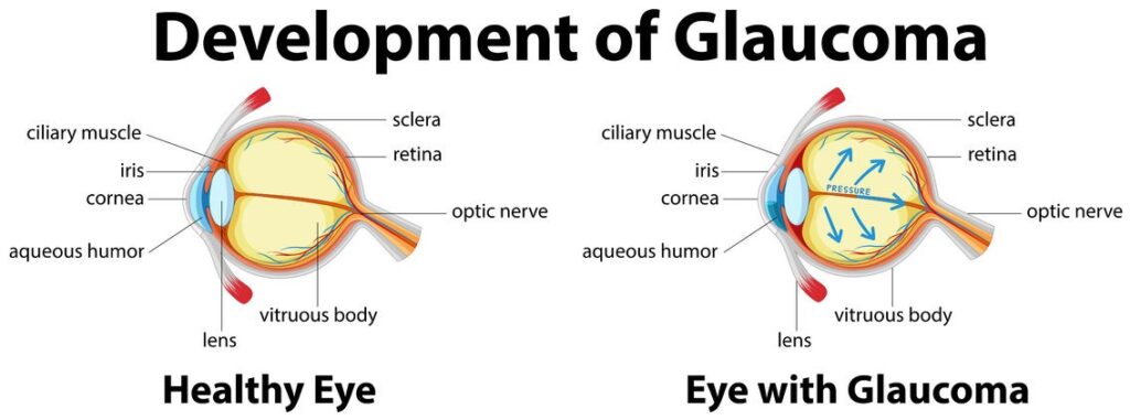
Glaucoma is often described as a condition of high intraocular pressure (IOP), or pressure inside the eye, leading to damage of the optic nerve. However, some cases of glaucoma are Normal-Tension Glaucoma (NTG), with normal levels of pressure on the optic nerve.  Treatment of glaucoma involve medication, surgery, or a combination.

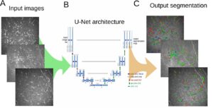
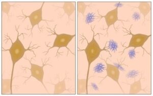
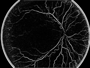
When we try to estimate how severe is a case of glaucoma, we have to look at the main area featuring the high damage of the pressure, which is the optic nerve. In that area, we notice a loss of retinal ganglion cells, photoreceptor nerve cells needed to transform light rays into electrical impulses, sent to the brain via the optic nerve. Their survival is necessary, but high pressure causes their death. In addition, we also observe a thinning of the Retinal Nerve Fiber Layer (RNFL): the choice way to assess these conditions for an accurate glaucoma detection is through cross-sectional 3D images taken by Optical Coherence Tomography (OCT) images or alternatively by color fundus images. OCT retinal images are very precise and enable our algorithms to make accurate measurements. The optic nerve is usually easy to detect, following the concentration of all blood vessels around its circular form. In addition to a possible glaucoma detection, all depressions on the surface of the retina are also detected.

Quantitative measurements are given following the deviation of tissues from the norm and the thickness of the Retinal Nerve Fiber Layer is measured after segmenting the different layers. This thickness is a precious and objective indicator of the status of the glaucoma.

Glaucoma Detection using deep learning

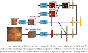
In a practical example using fundus color images, an algorithm detects the optical disc, which is the visible section of the optic nerve. Within that disc, a brighter area is found called the cup: when the cup-to-disc (C/D) ratio is larger than 0.3, expert ophthalmologists suspect a probable condition of glaucoma. Deep learning algorithms can help provide this assessment in an automated way using region of interest (the optical disc) and providing a probability for glaucoma, which the physician can use to support his diagnosis and treatment decisions.

Engineers at RSIP Vision have developed a very strong knowledge of the eye and of deep learning techniques, the combination of which enables us to help the medical experts take the most appropriate decisions for the patient, like we do in glaucoma detection and in many other ophthalmology tasks.

Share

Share on linkedin
Share on twitter
Share on facebook

Main Field

Ophthalmology

RSIP Vision has developed countless projects in the field of ophthalmology for its clients. Computer vision with sophisticated ophthalmic imaging, measurement techniques and AI can yield better results: great precision, accurate diagnosis and best interventional treatment for many pathologies.

View Ophthalmology

Categories

  • Ophthalmology, RSIP Vision Learns

Related Content

Zoom-in-Net

Deep Learning in Ophthalmology

Classification and Segmentation of Dendritic cells

Classification and Segmentation of Dendritic Cells

Alzheimer's Disease - AD

Degenerative Diseases Detection in the Eye

Temporary pediatric strabismus in newborn baby

Image processing for pediatric strabismus

ROP - Vessel tortuosity in Retinopathy of Prematurity

ROP: Retinopathy of Prematurity

Eyelid Drooping - MRD1 and MRD2

Eyelid Drooping – Blepharoptosis

Show all

RSIP Vision

Field-tested software solutions and custom R&D, to power your next medical products with innovative AI and image analysis capabilities.

Read more about us

Get in touch

Please fill the following form and our experts will be happy to reply to you soon

Recent News

PR – Intra-op Virtual Measurements in Laparoscopic and Robotic-Assisted Surgeries

PR – Non-Invasive Planning of Coronary Intervention

PR – Bladder Panorama Generator and Sparse Reconstruction Tool

PR – Registration Module for Orthopedic Surgery

All news
Upcoming Events
Stay informed for our next events
Subscribe to Our Magazines

Subscribe now and receive the Computer Vision News Magazine every month to your mailbox

 
Subscribe for free
Follow us
Linkedin Twitter Facebook Youtube

contact@rsipvision.com

Terms of Use

Privacy Policy

© All rights reserved to RSIP Vision 2021

Created by Shmulik

  • Our Work
    • title-1
      • Ophthalmology
      • Uncategorized
      • Ophthalmology
      • Pulmonology
      • Cardiology
      • Orthopedics
    • Title-2
      • Orthopedics
  • Success Stories
  • Insights
  • The company