Skip to content
  • Our Work
    • Fields
      • Cardiology
      • ENT
      • Gastro
      • Orthopedics
      • Ophthalmology
      • Pulmonology
      • Surgical Intelligence
      • Surgical Robotics
      • Urology
      • Other
    • Modalities
      • Endoscopy
      • Medical Segmentation
      • Microscopy
      • Ultrasound
  • Success Stories
  • Insights
    • Magazine
    • Upcoming Events
    • Webinars
    • Meetups
    • News
    • Blog
  • The company
    • About us
    • Careers
Menu
  • Our Work
    • Fields
      • Cardiology
      • ENT
      • Gastro
      • Orthopedics
      • Ophthalmology
      • Pulmonology
      • Surgical Intelligence
      • Surgical Robotics
      • Urology
      • Other
    • Modalities
      • Endoscopy
      • Medical Segmentation
      • Microscopy
      • Ultrasound
  • Success Stories
  • Insights
    • Magazine
    • Upcoming Events
    • Webinars
    • Meetups
    • News
    • Blog
  • The company
    • About us
    • Careers
Contact

Cardiac Left Ventricle Segmentation

Cardiac MRI is one of the technologies used for assessing the condition of patients which suffer or might suffer from a heart disease like cardiac masses and thrombi, pericardial abnormalities, cardiomyopathy, arrhythmia and coronary, aortic and/or different kinds of congenital cardiac diseases. Cardiac MRI is minimally invasive, does not involve radiation and it generally delivers excellent images for diagnostics.
Cardiac MRI with Left Ventricle
Cardiac MRI – chest cavity and heart
Of the four cardiac chambers which compose the heart, the left ventricle is quite often analyzed because its distinctive function (sending oxygenated blood out to distant tissue in the whole body) is vital for regular activity.  However, manual segmentation of the left ventricle is time-consuming and tiresome; it also shows highly inconsistent results between different observers. Thus, a fully or semi-automatic left ventricle segmentation is highly desirable.
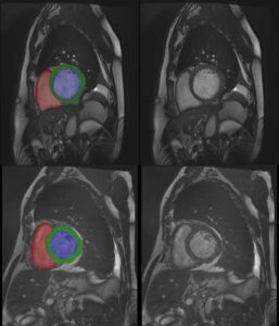
However, also automatic left ventricle segmentation (LV segmentation) faces challenging difficulties, like locating the left ventricle and overcoming the lack of edge information. We recommend a method to provide fast, accurate and fully automatic location and segmentation of the left ventricle, which takes into account also papillary and trabecular muscles, as well as identifies endocardial and epicardial contours in all the cardiac phases.

Left Ventricle Segmentation
Left ventricle segmentation

One of the main advantages of our recommended method is that it does not require any hand-made initialization, either by drawing manual contours or by adhering to a prior statistical heart shape model: the location of the left ventricle blood pool is automatically identified in the end diastole phase by the way of a roundness metric, under the assumptions that: the region of interest (the heart) is approximately in the middle of the image; that left and right ventricles’ blood pool have different circularity; and that blood has brighter signal intensity than the heart muscle. Another assumption is that the central part of the left ventricle blood pool does not move much across image slices and cardiac phases, which enables us to subsequently locate the contours of the blood pool, the papillary muscles (a small muscle within the ventricle that anchors the heart valves) and the trabeculations (pieces of muscle that extend into the ventricle). This is a crucial step, though it is sometimes neglected in other techniques of heart segmentation, since papillary muscles and trabeculations have a substantial impact on the measure of left ventricle volume, mass and ejection fraction1.
A region growing algorithm is used, after the image is converted to binary. Pixels are added in accordance with the intensity criterion (minimal difference in brightness). Image holes are then filled via morphological operations. Contours are finally smoothed with the help of a Fast Fourier Transform (FFT) technique, which removes all data points that are far from most other points of the dataset, without changing the overall shape.
The proposed method provides very robust input for daily clinical application, in line with expert-found values. Its only limitation to date is in the area of detecting contours in hypertrophic subjects. Future research will introduce additional constraints to take care also of this difficulty.

1 The health of the left ventricle can be (among other means) by quantitatively measuring its ejection fraction, stroke volume and ventricular mass, all critical parameters for cardiac diagnosis. Ejection fraction quantifies the percentage volume of blood spread out of the left ventricle in a given cardiac cycle. Stroke volume is the amount of blood pumped out of the left ventricle to the body.
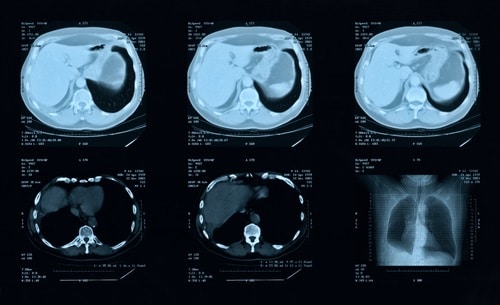
Share

Share on linkedin
Share on twitter
Share on facebook

Main Field

Cardiology

RSIP Vision’s advanced algorithms and image processing software (together with proper machines and devices) are used by physicians and researchers to develop and perform preventive and interventional cardiovascular procedures: that's our contribution to Artificial Intelligence in cardiology. Our software provides information about the patient’s cardiovascular system in a non-invasive manner, helps physicians ensure accuracy during complex operations, raises the level of patient safety through limiting his/her exposure to radiation, and aids diagnosis. We come to the office to write algorithms that save lives. In this section about computer vision in cardiology you can discover some of the ways we do it.

View Cardiology

Categories

  • Cardiology, RSIP Vision Learns

Related Content

PR – Non-Invasive Planning of Coronary Intervention

AI in Cardiac CT angiography

Coronary Arteries Segmentation

Great Vessels Segmentation with Deep Learning

Cardiac Motion Correction

Deep Learning in Cardiology

Cardiac MRI 3D automatic segmentation

3D Cardiac MRI automatic segmentation

PR – Non-Invasive Planning of Coronary Intervention

AI in Cardiac CT angiography

Coronary Arteries Segmentation

Great Vessels Segmentation with Deep Learning

Cardiac Motion Correction

Deep Learning in Cardiology

Cardiac MRI 3D automatic segmentation

3D Cardiac MRI automatic segmentation

Show all

RSIP Vision

Field-tested software solutions and custom R&D, to power your next medical products with innovative AI and image analysis capabilities.

Read more about us

Get in touch

Please fill the following form and our experts will be happy to reply to you soon

Recent News

PR – Intra-op Virtual Measurements in Laparoscopic and Robotic-Assisted Surgeries

PR – Non-Invasive Planning of Coronary Intervention

PR – Bladder Panorama Generator and Sparse Reconstruction Tool

PR – Registration Module for Orthopedic Surgery

All news
Upcoming Events
Stay informed for our next events
Subscribe to Our Magazines

Subscribe now and receive the Computer Vision News Magazine every month to your mailbox

 
Subscribe for free
Follow us
Linkedin Twitter Facebook Youtube

contact@rsipvision.com

Terms of Use

Privacy Policy

© All rights reserved to RSIP Vision 2021

Created by Shmulik

  • Our Work
    • title-1
      • Ophthalmology
      • Uncategorized
      • Ophthalmology
      • Pulmonology
      • Cardiology
      • Orthopedics
    • Title-2
      • Orthopedics
  • Success Stories
  • Insights
  • The company