Skip to content
  • Our Work
    • Fields
      • Cardiology
      • ENT
      • Gastro
      • Orthopedics
      • Ophthalmology
      • Pulmonology
      • Surgical Intelligence
      • Surgical Robotics
      • Urology
      • Other
    • Modalities
      • Endoscopy
      • Medical Segmentation
      • Microscopy
      • Ultrasound
  • Success Stories
  • Insights
    • Magazine
    • Upcoming Events
    • Webinars
    • Meetups
    • News
    • Blog
  • The company
    • About us
    • Careers
Menu
  • Our Work
    • Fields
      • Cardiology
      • ENT
      • Gastro
      • Orthopedics
      • Ophthalmology
      • Pulmonology
      • Surgical Intelligence
      • Surgical Robotics
      • Urology
      • Other
    • Modalities
      • Endoscopy
      • Medical Segmentation
      • Microscopy
      • Ultrasound
  • Success Stories
  • Insights
    • Magazine
    • Upcoming Events
    • Webinars
    • Meetups
    • News
    • Blog
  • The company
    • About us
    • Careers
Contact

Retinal inner layers segmentation

OCT is the only method that can perform noninvasive imaging with non-ionizing radiation and offering relatively good resolution. That is why it has become a standard in clinical ophthalmology. Segmentation of anatomical and pathological structures in ophthalmic images is crucial for the diagnosis and study of ocular and other diseases; for example, patients may have quantitative abnormalities in different retinal layers in multiple sclerosis (MS), diabetes, Alzheimer’s, Parkinson’s, and glaucoma. To analyze the effects of these pathologies on specific types of cells within the retina, it is necessary to accurately segment the different tissue layers that appear in retinal OCT images. This is the task of the retinal inner layers segmentation.

Spectral Domain Optical Coherence Tomography (SD-OCT)

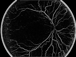
Since manual segmentation of retinal layers is tedious, time-consuming and subjective, it is necessary that such segmentation be automated. Automated detection of retinal layers is best done on OCT images but it has to take into account a few challenges. The following is how RSIP Vision engineers analyze the problem and the options to solve it.

The reasonably good resolution of OCT images enables to distinguish the different retinal layers. When we need additional information about these layers, we would like to have an automated system to segment them.

The most common segmentation approaches are: intensity-based B-scan analysis (based on intensity variations and gradients), active contour approaches, pattern recognition analysis methods, and segmentation methods using 2D or 3D graphs constructed from OCT datasets.

Before beginning the segmentation it is essential to do some preprocessing of the data using intensity and spatial normalization, and in certain methods also flattening the retina. The first step in this automated detection of retinal layers will require an initial segmentation of the ILM-to-RPE complex, the outer borders of the retina. ILM is the Inner Limiting Membrane of the retina, while the RPE is the Retinal Pigment Epithelium. Distance between these two opposite edges is usually considered the most common measurement of retinal thickness. Their identification is quite straightforward, since RPE is bright and ILM is the last layer before the background. However, segmentation of this area presents at least one challenge: trusting brightness alone can be problematic in OCT images because of the high levels of noise, which can take the form of line discontinuation or other layers having bright reflections too; discontinuations are missing data in a particular zone of the layer and may be due to blood vessels obstructing the view to the area and sometimes because the tissue itself does not have enough texture to be distinguishable. Also, an eye affected by pathologies does not present as regular layers as a healthy eye. Curve interpolation is one method that can compensate on the missing data.

Detection of the inner layers of the retina is a problem of higher complexity than the retinal edges. Every layer has its own appearance and we expect to see them accordingly in the OCT image. Thus, it is possible to rely on statistical methods to identify each layer, following its structure, brightness and relative position in the series of layers or using texture analysis strategy.

Retinal inner layers segmentation techniques

The following step, the one that will deliver the final segmentation itself, needs to use an optimization method, since it is impossible to decide about the layer on the pixel level: only seeing the whole image gives the complete information needed.

Two methods commonly used to approach this area are the active contour approach and graph theory techniques like graph-cuts or Bellman-Ford. When we use active contour, we know a priori what specific layers we are looking for: what we need is to locate them in the image. To assist us, we have parametric weights that derivate from distances between pixels or differences between intensity values, for the optimization method: the inner energy term tends to make the graph smoother and stable; the outer energy term drives the active contour towards the significant features of the image, like boundaries. Using this algorithm iteratively for all layers brings us closer to the final segmentation.

A more advanced approach constructs a geometric graph to simultaneously segment all boundaries in a 3-D OCT volume. Unlike previously presented approaches, it takes into account the interaction of neighboring boundaries to mutually restrict their relative positions.

Like many tasks in ophthalmology image processing, automated retinal inner layers segmentation needs deep knowledge of both the morphology of the eye and the algorithmic techniques to choose from in order to get an accurate and fast segmentation. RSIP Vision’s engineers have a solid experience in both fields. Contact our consultants to check if and how we can help you on your computer vision projects.

Share

Share on linkedin
Share on twitter
Share on facebook

Main Field

Ophthalmology

RSIP Vision has developed countless projects in the field of ophthalmology for its clients. Computer vision with sophisticated ophthalmic imaging, measurement techniques and AI can yield better results: great precision, accurate diagnosis and best interventional treatment for many pathologies.

View Ophthalmology

Categories

  • Ophthalmology, RSIP Vision Learns

Related Content

Zoom-in-Net

Deep Learning in Ophthalmology

Classification and Segmentation of Dendritic cells

Classification and Segmentation of Dendritic Cells

Alzheimer's Disease - AD

Degenerative Diseases Detection in the Eye

Temporary pediatric strabismus in newborn baby

Image processing for pediatric strabismus

ROP - Vessel tortuosity in Retinopathy of Prematurity

ROP: Retinopathy of Prematurity

Eyelid Drooping - MRD1 and MRD2

Eyelid Drooping – Blepharoptosis

Show all

RSIP Vision

Field-tested software solutions and custom R&D, to power your next medical products with innovative AI and image analysis capabilities.

Read more about us

Get in touch

Please fill the following form and our experts will be happy to reply to you soon

Recent News

PR – Intra-op Virtual Measurements in Laparoscopic and Robotic-Assisted Surgeries

PR – Non-Invasive Planning of Coronary Intervention

PR – Bladder Panorama Generator and Sparse Reconstruction Tool

PR – Registration Module for Orthopedic Surgery

All news
Upcoming Events
Stay informed for our next events
Subscribe to Our Magazines

Subscribe now and receive the Computer Vision News Magazine every month to your mailbox

 
Subscribe for free
Follow us
Linkedin Twitter Facebook Youtube

contact@rsipvision.com

Terms of Use

Privacy Policy

© All rights reserved to RSIP Vision 2021

Created by Shmulik

  • Our Work
    • title-1
      • Ophthalmology
      • Uncategorized
      • Ophthalmology
      • Pulmonology
      • Cardiology
      • Orthopedics
    • Title-2
      • Orthopedics
  • Success Stories
  • Insights
  • The company