Skip to content
  • Our Work
    • Fields
      • Cardiology
      • ENT
      • Gastro
      • Orthopedics
      • Ophthalmology
      • Pulmonology
      • Surgical Intelligence
      • Surgical Robotics
      • Urology
      • Other
    • Modalities
      • Endoscopy
      • Medical Segmentation
      • Microscopy
      • Ultrasound
  • Success Stories
  • Insights
    • Magazine
    • Upcoming Events
    • Webinars
    • Meetups
    • News
    • Blog
  • The company
    • About us
    • Careers
Menu
  • Our Work
    • Fields
      • Cardiology
      • ENT
      • Gastro
      • Orthopedics
      • Ophthalmology
      • Pulmonology
      • Surgical Intelligence
      • Surgical Robotics
      • Urology
      • Other
    • Modalities
      • Endoscopy
      • Medical Segmentation
      • Microscopy
      • Ultrasound
  • Success Stories
  • Insights
    • Magazine
    • Upcoming Events
    • Webinars
    • Meetups
    • News
    • Blog
  • The company
    • About us
    • Careers
Contact

Right Atrium Measurement with Ultrasound

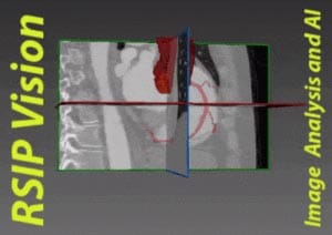
Right Atrium Measurement in Ultrasound Videos


Atrial fibrillation is an irregular rhythmic beating of the heart associated with coronary heart disease, high blood pressure and blood clots. Detection and tracking of conditions leading to it is routinely done by observing the heart using noninvasive ultrasound imaging (echocardiogram). Observations are particularly focused on the performance of the right atrium.

Irregularity in the volume of the right atrium indicates the presence of severe disease. Enlargement of the right atrium might result from volume or pressure load. Detection and estimation of the severity of the pressure can be performed by measuring the size and shape of the vena cava, entering and exiting the right atrium.

The right atrium is a hollow chamber in the upper right side of the heart, responsible for pumping the blood. Deoxygenated blood enters the right atrium through veins and from there it is pushed to the right ventricle on its way to be oxygenated in the lungs.

Changes in the volume and shape of the right atrium indicates its flexibility to contract. Viewing and tracking these changes allows physicians to estimate whether the heart is functioning correctly. The shape and borders of the right atria indicates the volume pressure.

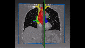
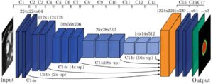
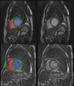
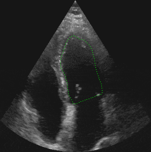
Automatic detection and measurement of the right atrium aids physicians to estimate the severity of pressure load or the existence of other pathologies. It is therefore key to provide the medical profession with a precise right atrium measurement tool. For this end, RSIP Vision has constructed software which automatically measures the right atrium. The videos we have analyzed are in 4 chamber view, in which the heart shape changes throughout its cycle. The dynamic changes complicate the detection and tracking. RSIP Vision has overcome the challenges of dynamic heart view using advanced image processing methods to track deformable objects. Coupled with machine learning procedures we have produced an algorithm to accurately measure the shape and volume of the right atrium.

Cardiovascular Ultrasound software at work

The significance of fast automatic detection of pathologies in right atrium is emphasized with the increased availability of bedside ultrasound used in emergency rooms to detect pathologies and manage patients. Our algorithms can operate in real-time in those machines to give valuable information in routine procedures or emergencies.

Share

Share on linkedin
Share on twitter
Share on facebook

Main Field

Cardiology

RSIP Vision’s advanced algorithms and image processing software (together with proper machines and devices) are used by physicians and researchers to develop and perform preventive and interventional cardiovascular procedures: that's our contribution to Artificial Intelligence in cardiology. Our software provides information about the patient’s cardiovascular system in a non-invasive manner, helps physicians ensure accuracy during complex operations, raises the level of patient safety through limiting his/her exposure to radiation, and aids diagnosis. We come to the office to write algorithms that save lives. In this section about computer vision in cardiology you can discover some of the ways we do it.

View Cardiology

Categories

  • Cardiology, RSIP Vision Learns

Related Content

PR – Non-Invasive Planning of Coronary Intervention

AI in Cardiac CT angiography

Coronary Arteries Segmentation

Great Vessels Segmentation with Deep Learning

Cardiac Motion Correction

Deep Learning in Cardiology

Cardiac MRI 3D automatic segmentation

3D Cardiac MRI automatic segmentation

PR – Non-Invasive Planning of Coronary Intervention

AI in Cardiac CT angiography

Coronary Arteries Segmentation

Great Vessels Segmentation with Deep Learning

Cardiac Motion Correction

Deep Learning in Cardiology

Cardiac MRI 3D automatic segmentation

3D Cardiac MRI automatic segmentation

Show all

RSIP Vision

Field-tested software solutions and custom R&D, to power your next medical products with innovative AI and image analysis capabilities.

Read more about us

Get in touch

Please fill the following form and our experts will be happy to reply to you soon

Recent News

PR – Intra-op Virtual Measurements in Laparoscopic and Robotic-Assisted Surgeries

PR – Non-Invasive Planning of Coronary Intervention

PR – Bladder Panorama Generator and Sparse Reconstruction Tool

PR – Registration Module for Orthopedic Surgery

All news
Upcoming Events
Stay informed for our next events
Subscribe to Our Magazines

Subscribe now and receive the Computer Vision News Magazine every month to your mailbox

 
Subscribe for free
Follow us
Linkedin Twitter Facebook Youtube

contact@rsipvision.com

Terms of Use

Privacy Policy

© All rights reserved to RSIP Vision 2021

Created by Shmulik

  • Our Work
    • title-1
      • Ophthalmology
      • Uncategorized
      • Ophthalmology
      • Pulmonology
      • Cardiology
      • Orthopedics
    • Title-2
      • Orthopedics
  • Success Stories
  • Insights
  • The company