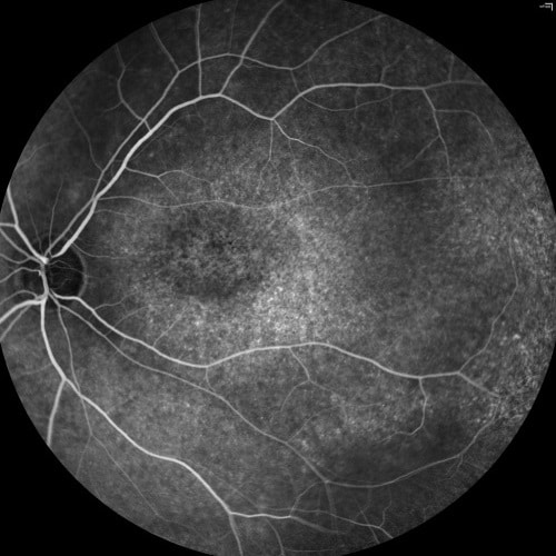
Stabilization in fluorescein angiography
This eye angiogram test is very efficient in finding leaks in veins and arteries. In order to locate with precision the area where that happens, it is necessary to stabilize the video, because during the test eyes still move and there might also be very quick saccades. When no correction is done, Saccadic eye motions will be visible in OCT angiogram. This is why stabilization in fluorescein angiography video clips is a crucial task in computer vision for ophthalmology.
RSIP Vision has developed a stabilizer for this video clip, so that the ophthalmologist can see a very stable eye and within a few moments all the colors are changing according to the tone of circulating blood.
Stabilization in fluorescein angiography is not devoid of challenges: one can try to use correlation in many of the registration / stabilization tasks in image processing. The advantage of correlation is that it has very good implementation in hardware and even in GPU, so that you can do it very fast with very reliable results. However, since the entire image is changing, during those moments the appearance of the blood vessels varies accordingly and much more stable measures are required in order to help us find points of interest which are still in the image, even when it changes its appearance.
One of the solutions might be a mutual information strategy where instead of looking at the complete identity between the values in the different images, we look at information changes in the different parts of the image and we use that to give a score to the matching of the images and perform the registration.
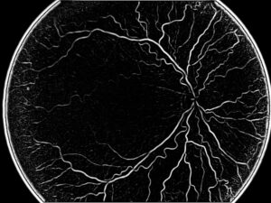
A different strategy might use key points, which would remain stable under the different circumstances, like junction points and bifurcation points. We can also use a third system, that will look at the derivative images focusing on the boundaries of the vessels rather than on the raw information of the pixel values. We have thus three different solutions, having different advantages and applications. RSIP Vision engineers have a very rich experience that enables them to find the optimal method to use in each situation.
 Ophthalmology