Skip to content
  • Our Work
    • Fields
      • Cardiology
      • ENT
      • Gastro
      • Orthopedics
      • Ophthalmology
      • Pulmonology
      • Surgical Intelligence
      • Surgical Robotics
      • Urology
      • Other
    • Modalities
      • Endoscopy
      • Medical Segmentation
      • Microscopy
      • Ultrasound
  • Success Stories
  • Insights
    • Magazine
    • Upcoming Events
    • Webinars
    • Meetups
    • News
    • Blog
  • The company
    • About us
    • Careers
Menu
  • Our Work
    • Fields
      • Cardiology
      • ENT
      • Gastro
      • Orthopedics
      • Ophthalmology
      • Pulmonology
      • Surgical Intelligence
      • Surgical Robotics
      • Urology
      • Other
    • Modalities
      • Endoscopy
      • Medical Segmentation
      • Microscopy
      • Ultrasound
  • Success Stories
  • Insights
    • Magazine
    • Upcoming Events
    • Webinars
    • Meetups
    • News
    • Blog
  • The company
    • About us
    • Careers
Contact

Multi-modal registration of retina images

Multi-modal registration of retina images is a process by which pictures taken with different modalities and/or at different times are registered. This is done principally when tracking some kind of condition concerning the patient, e.g. the evolution of a disease.
Multi-Modal Image Registration
Software by RSIP Vision automatically performs registration of images from different modalities (different imaging devices or technologies).

One of the things that ophthalmologist want to do is comparing the progress of a disease or the results of a treatment administered to try and solve that condition. In particular, physicians want to be able to compare images taken with different devices (representing different modalities): that allows to examine the same area from different points of view. In fact, some modalities are better in emphasizing specific pathologies, while others are better equipped to examine other aspects of the patient’s condition. Putting one image over the other enables the eye health professional to switch and see how the phenomena looks under different imaging techniques.

Image analysis modalities in ophthalmology:

The set of modalities used for retinal imaging is not the same as the set used for other body parts (though it is parallel to it): MRI, CT, X-Ray and even Ultrasound. The modalities used in eye care imaging are: regular fundus images, fluorescein angiography, Indocyanine Green Chorioangiography (ICG), Optical Coherence Tomography (OCT), Fundus Autofluorescence (FAF) imaging: each of these modalities reveals a different aspect about the eye. For instances, slices of OCT can show specific points in the retina that need to be examined.

Stabilization of saccades (rapid movements of the eye) is another very important procedure which is done with fluorescein angiography or with ICG imaging: by that process, you can see the same area in stabilized images, so that you can observe its changes over time.


Algorithmic techniques for Multi-Modality Registration

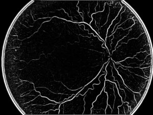
The appearance of arteries changes very significantly when using different modalities. The same retinal arteries which look dark red in fundus imaging will turn from white to black in fluorescein imaging. This excludes the use of simple correlation methods for this multi-modal registration task. Usually, one can use mutual information techniques or some markers, which can be identified by Scale Invariant Feature Transform (SIFT) or Speeded Up Robust Features (SURF), two local feature detectors and descriptors. That will capture interest points like bifurcation or any other interesting zone that you can lock on. Images will then be overlaid one on another.

This is done together with noise reduction and other processing techniques for clearing detailed images. Other technologies might use some gradient image, which might be common to different modalities.


Importance for the patient’s electronically-stored health records:

This procedure is valuable for another important reason: in addition to provide crucial information to the doctor, it also proves very useful in front of advanced Electronic Medical Record (EMR) and Electronic Health Record (EHR) systems, which become not only a data and image repository, but also a tool for comparison and sophisticated uses, giving the physician better tools to assess progression or regression and understand the status of his or her patient’s pathologies.

Share

Share on linkedin
Share on twitter
Share on facebook

Main Field

Ophthalmology

RSIP Vision has developed countless projects in the field of ophthalmology for its clients. Computer vision with sophisticated ophthalmic imaging, measurement techniques and AI can yield better results: great precision, accurate diagnosis and best interventional treatment for many pathologies.

View Ophthalmology

Categories

  • Ophthalmology, RSIP Vision Learns

Related Content

Zoom-in-Net

Deep Learning in Ophthalmology

Classification and Segmentation of Dendritic cells

Classification and Segmentation of Dendritic Cells

Alzheimer's Disease - AD

Degenerative Diseases Detection in the Eye

Temporary pediatric strabismus in newborn baby

Image processing for pediatric strabismus

ROP - Vessel tortuosity in Retinopathy of Prematurity

ROP: Retinopathy of Prematurity

Eyelid Drooping - MRD1 and MRD2

Eyelid Drooping – Blepharoptosis

Show all

RSIP Vision

Field-tested software solutions and custom R&D, to power your next medical products with innovative AI and image analysis capabilities.

Read more about us

Get in touch

Please fill the following form and our experts will be happy to reply to you soon

Recent News

PR – Intra-op Virtual Measurements in Laparoscopic and Robotic-Assisted Surgeries

PR – Non-Invasive Planning of Coronary Intervention

PR – Bladder Panorama Generator and Sparse Reconstruction Tool

PR – Registration Module for Orthopedic Surgery

All news
Upcoming Events
Stay informed for our next events
Subscribe to Our Magazines

Subscribe now and receive the Computer Vision News Magazine every month to your mailbox

 
Subscribe for free
Follow us
Linkedin Twitter Facebook Youtube

contact@rsipvision.com

Terms of Use

Privacy Policy

© All rights reserved to RSIP Vision 2021

Created by Shmulik

  • Our Work
    • title-1
      • Ophthalmology
      • Uncategorized
      • Ophthalmology
      • Pulmonology
      • Cardiology
      • Orthopedics
    • Title-2
      • Orthopedics
  • Success Stories
  • Insights
  • The company