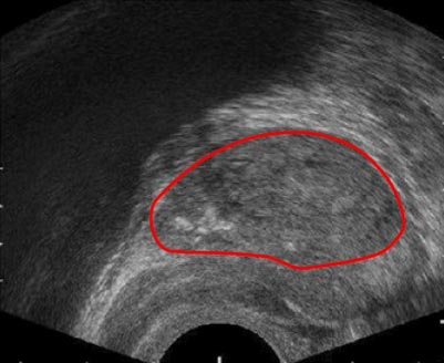
Image Analysis and AI for BPH
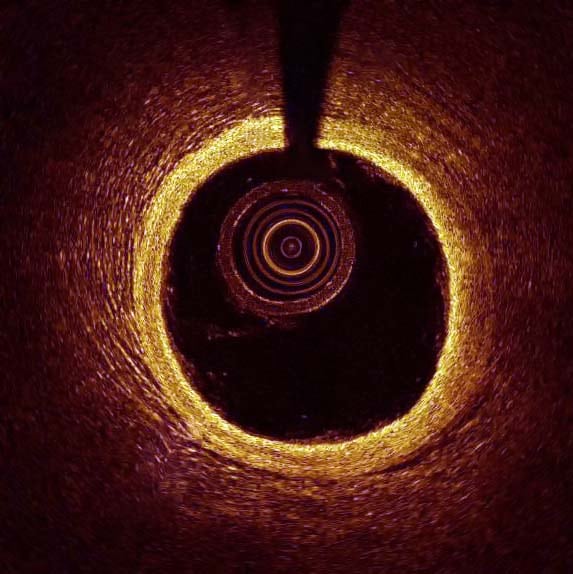
Endoscopic Procedures using Intravenous OCT
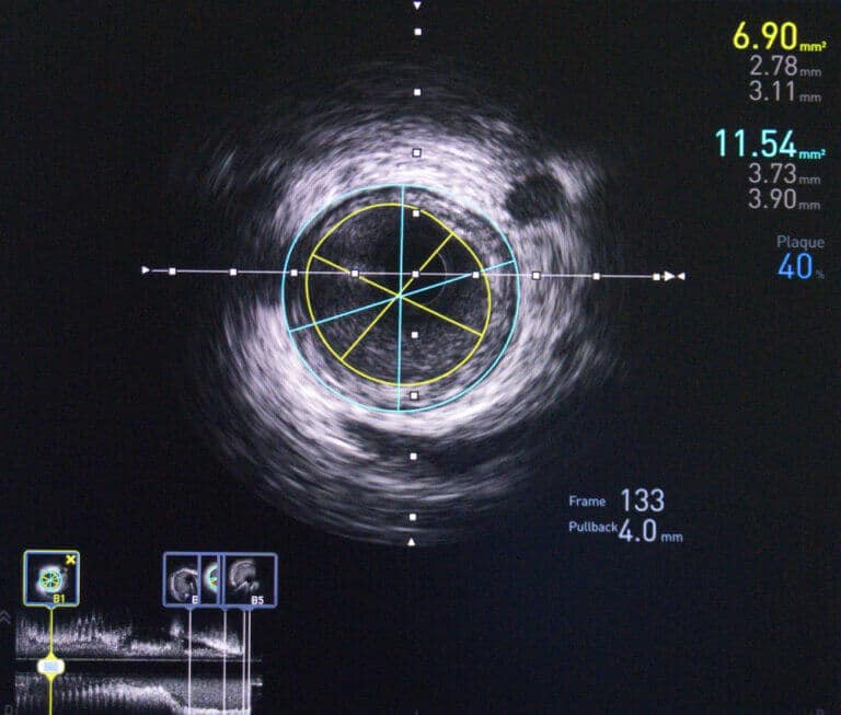
Using AI to Analyse Intravenous Ultrasound Images
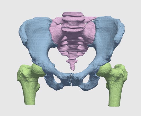
Segmentation in Orthopedics with Deep Learning
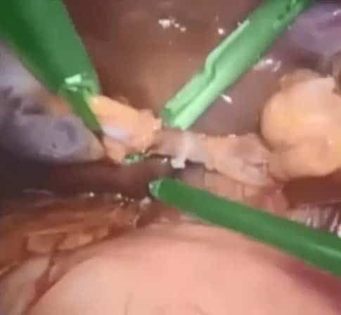
Surgical Instrument Segmentation
AI-based Surgical Workflow Analysis, another big step towards the future of robotic surgery
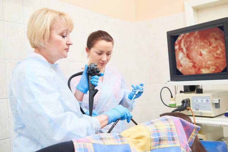
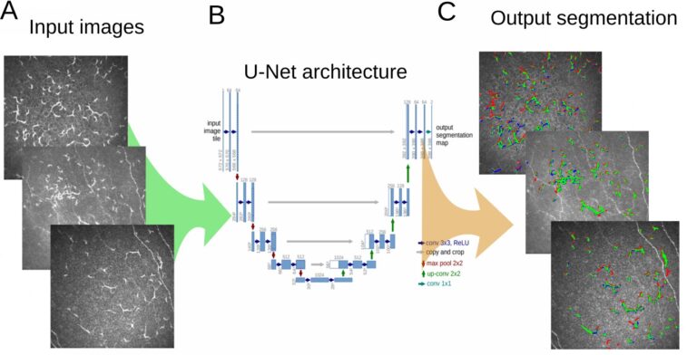
Challenges and AI Solutions for Endoscopy
As endoscopic and microscopic image processing, and surgical vision are evolving as necessary tools for computer assisted interventions (CAI), researchers have recognized the need for
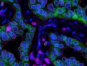
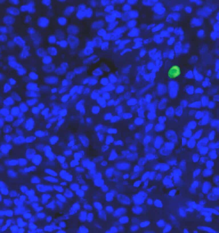
Multiplex IF Analysis
The use of deep learning for analysis of multiplex IF has allowed for a much greater accuracy level for the correct phenotypic classification of cells. When combined with RSIP Vision‘s advanced nuclear detection capability, it allows for the simultaneous analysis of multiple florescent markers on a cell by cell basis. This tool is well suited for multiple applications, especially when using multiple markers to characterize distinct cell populations such as in immune-oncology and IBD.
Circulating Tumor Cells (CTCs)
Circulating tumor cells (CTCs) are rare cancer cells that originate from a tumor and then travel through the patient’s blood or lymphatic system. CTCs have
Coronary Arteries Segmentation
Coronary artery disease (CAD) or ischemic heart disease (IHD) has become one of the most common causes of morbidity and mortality worldwide. Patients who suffer
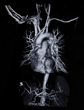
Great Vessels Segmentation with Deep Learning
The great vessels conduct blood to and from the heart. These vessels include the aorta, superior and inferior vena cava, pulmonary arteries and pulmonary veins.
Middle and Inner Ear Segmentation with Deep Learning
Ear pathologies are common in all age groups, and are one of the leading causes for visiting a doctor. In most cases, proper diagnosis can
Larynx Segmentation with Deep Learning
The larynx, also known as the voice box, is a triangular structure in charge of important functions including breathing, voice production and supplying protection to
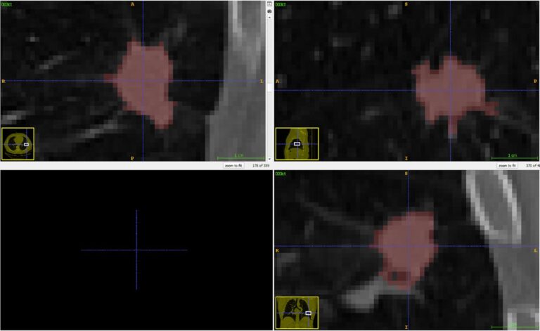
Lymph Node Segmentation Module
Lymph nodes are routinely examined and assessed during physical examination of patients in a clinic or hospital setting. Enlarged lymph nodes can be indicators of
Brain Ventricles Segmentation with Deep Learning
Brain Hemorrhage Segmentation with Deep Learning
Detection and Tracking of Tumors
RSIP Vision’s oncology software combines detection of lesions and tumors in the human body with tracking those findings along CT scans performed during the research: in particular lung, lymph nodes and liver. These tools enable a quick and accurate assessment of the efficacy of the new treatment.

Automated RECIST Measurement
The golden standard for measuring tumors is the RECIST score. RSIP Vision developed an automated module to accurately measure the RECIST score from CT scans as well as the exact 3D volume of the tumors. Changes in volume are a reliable measure of the progression or remission of the tumor, enabling to evaluate the responsiveness of the treatment in a relatively short time.

Intrusion Detection with Deep Learning
Detecting physical and virtual intrusions is a key process in ensuring information and property security. Physical intrusion detection refers to all attempts at break-ins to
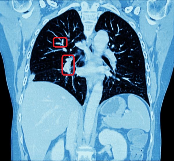
Chest CT Scan Analysis with Deep Learning
Chest radiography, with modalities such as X-Ray and CT, is now the common practice for the detection and analysis of the progression of lung tumors,
Object Tracking at High fps
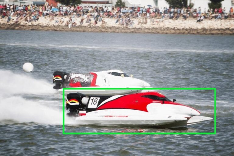
Object tracking in video sequences is a classical challenge in computer vision, which finds applications in nearly all domains of the industry: from assembly line
High Resolution Image Reconstruction
Recovering a high-resolution (HR) image from a low resolution one is a classical problem in computer vision for which many algorithms have been developed to
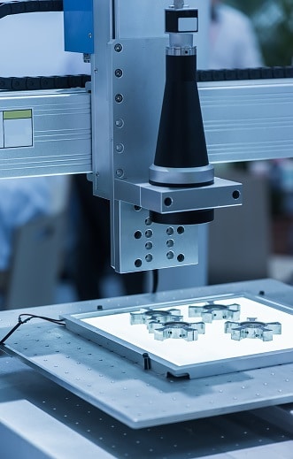
Automated Defect Inspection Using Deep Learning
Deep Learning in Cardiology
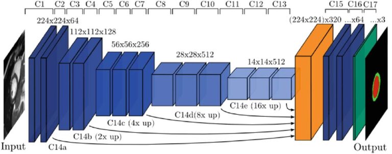
1.1 Segmentation tasks [10] suggest a new fully convolutional network architecture for the task of cardiovascular MRI segmentation. The architecture is based on the idea
Deep Learning in Pulmonology
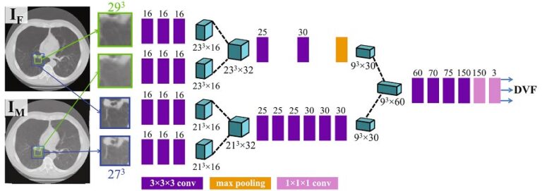
Deep learning has been successfully applied in various applications in pulmonary imaging, including CT registration, airway mapping, real time catheter navigation, and pulmonary nodule detection.
Deep Learning in Ophthalmology
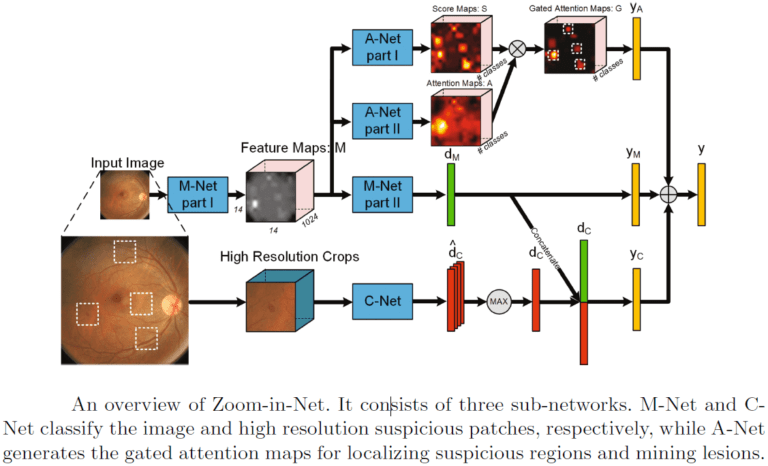
Recent works suggest novel deep learning tools for detection, segmentation and characterization of eye disorders. Accurate segmentation of retinal fundus lesions and anomalies in imaging
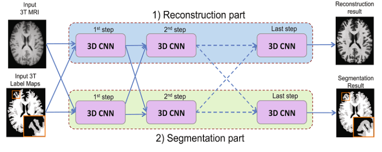
Deep Learning in Brain Imaging
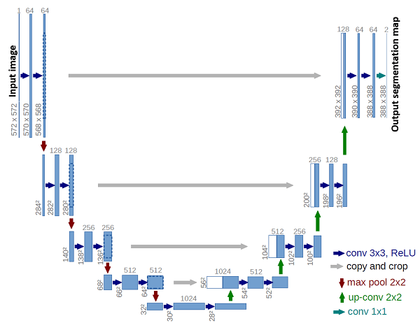
Deep Learning in Medical Imaging
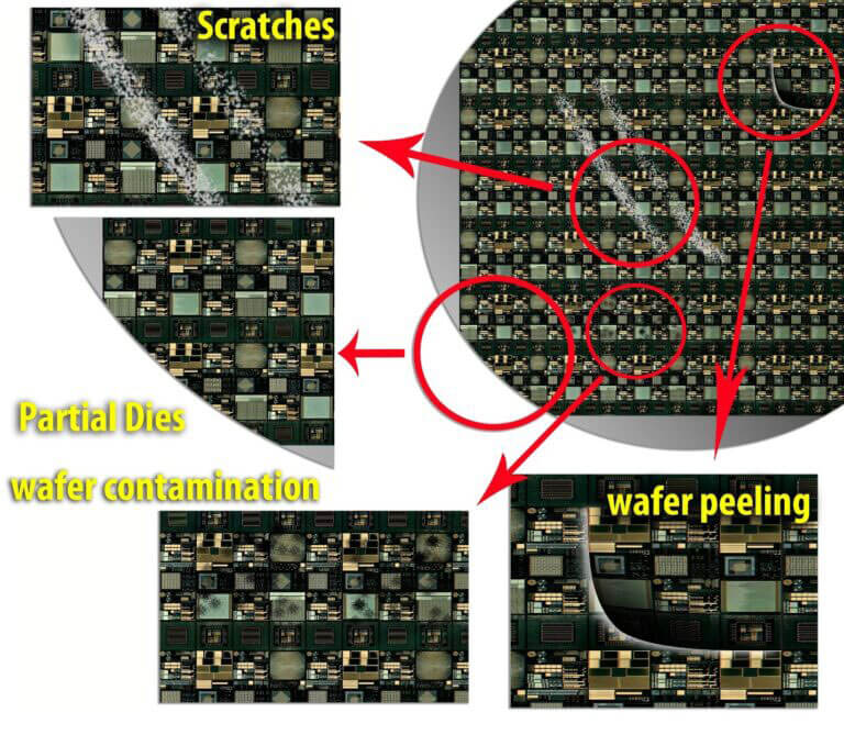
Wafer Macro Defects Detection and Classification
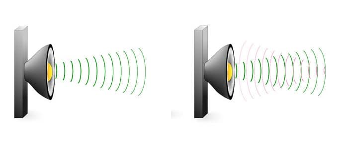
Echo Cancellation Using Deep Learning
Complete cancellation of returned acoustic echo signal is still an unresolved issue in signal processing. When a signal from a speaker in one end of
Classification and Segmentation of Dendritic Cells
Dry eye disease (DED) is one of the most common ophthalmic disorders. Inflammation of the ocular surface is controlled by corneal antigen-presenting cells called dendritic

Fingerprint Segmentation Using Deep Learning
Automatic fingerprint recognition systems are based on the extraction of features from scanned fingerprint image. A successful preprocessing of the scan is an important first
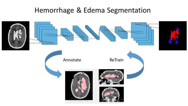
Intracranial Hemorrhage and Edema Segmentation
An intracranial hemorrhage (ICH) is a condition in which a blood vessel erupts inside the brain, causing internal bleeding. If not treated correctly and immediately,
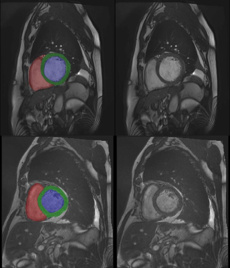
3D Cardiac MRI automatic segmentation
Object Detection Methods for Robots
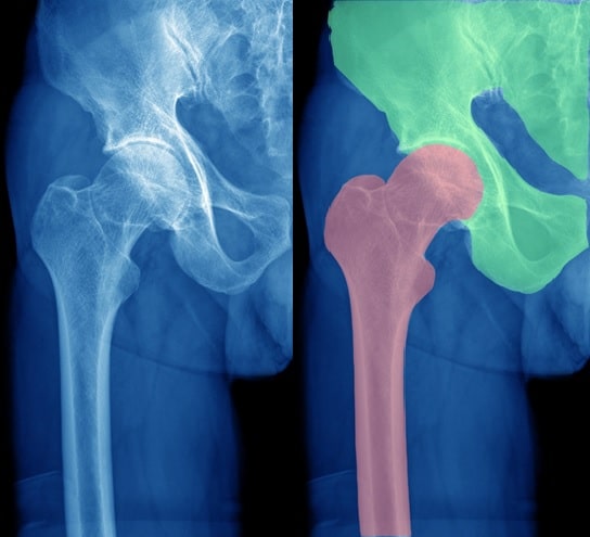
CT Segmentation in orthopedic surgery
CT image segmentation is a typical phase of orthopedic surgeries in which a visualization system is called to visually support the surgeon’s task. This system
Coronary CT Angiography with Deep Learning

Automatic semantic tagging of images

Detecting Mitosis Using Deep Neural Networks
Lungs tumors and nodules segmentation with Deep Learning
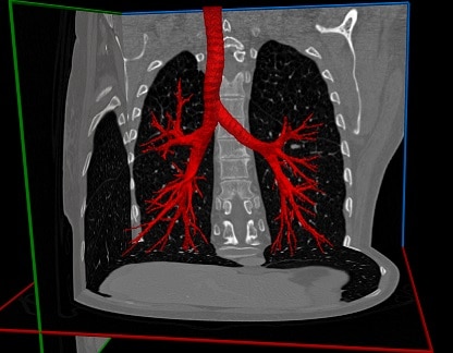
Airways segmentation with Deep Learning
Kidney Segmentation

Vessel Segmentation Using Deep Learning
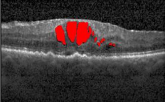
Finding Cysts, Part Five: Final Detection
Automatic Detection of Macular Cysts

Defining the Borders within Computer Vision
What’s the Difference between Computer Vision, Image Processing and Machine Learning? In this page, you will learn about Machine Vision, Computer Vision and Image Processing. If you
Reflections on CVPR 2015
Reflections on CVPR 2015 One of our colleagues, Dr. Micha Feigin, presents his thoughts on this year’s Computer Vision & Pattern Recognition Conference: Coming back
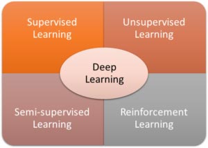
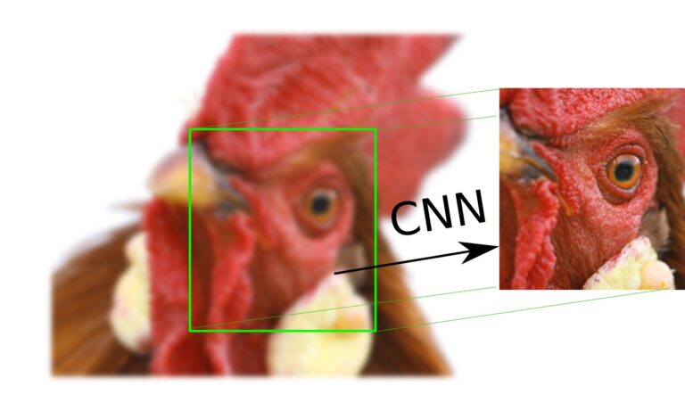
Exploring Deep Learning & CNNs
Deep Learning and Convolutional Neural Networks: RSIP Vision Blogs In this page, you will learn about Computer Vision, Machine Vision and Image Processing. If